
Profile of Specialty Focused Journal Supplements
| Classics and Revisits in Scientific Cardiology.CRS[Card.]
A focused review of the Cardiac Anatomy Angina Pectoris and Acute Chest pain of Cardiac origin |
Classics and Revisits in Scientific Dermatology CRS [Derm.] A directed anatomical study for the dermatological structures |
| Classics and Revisits in Scientific Endocrinology CRS [End.]
|
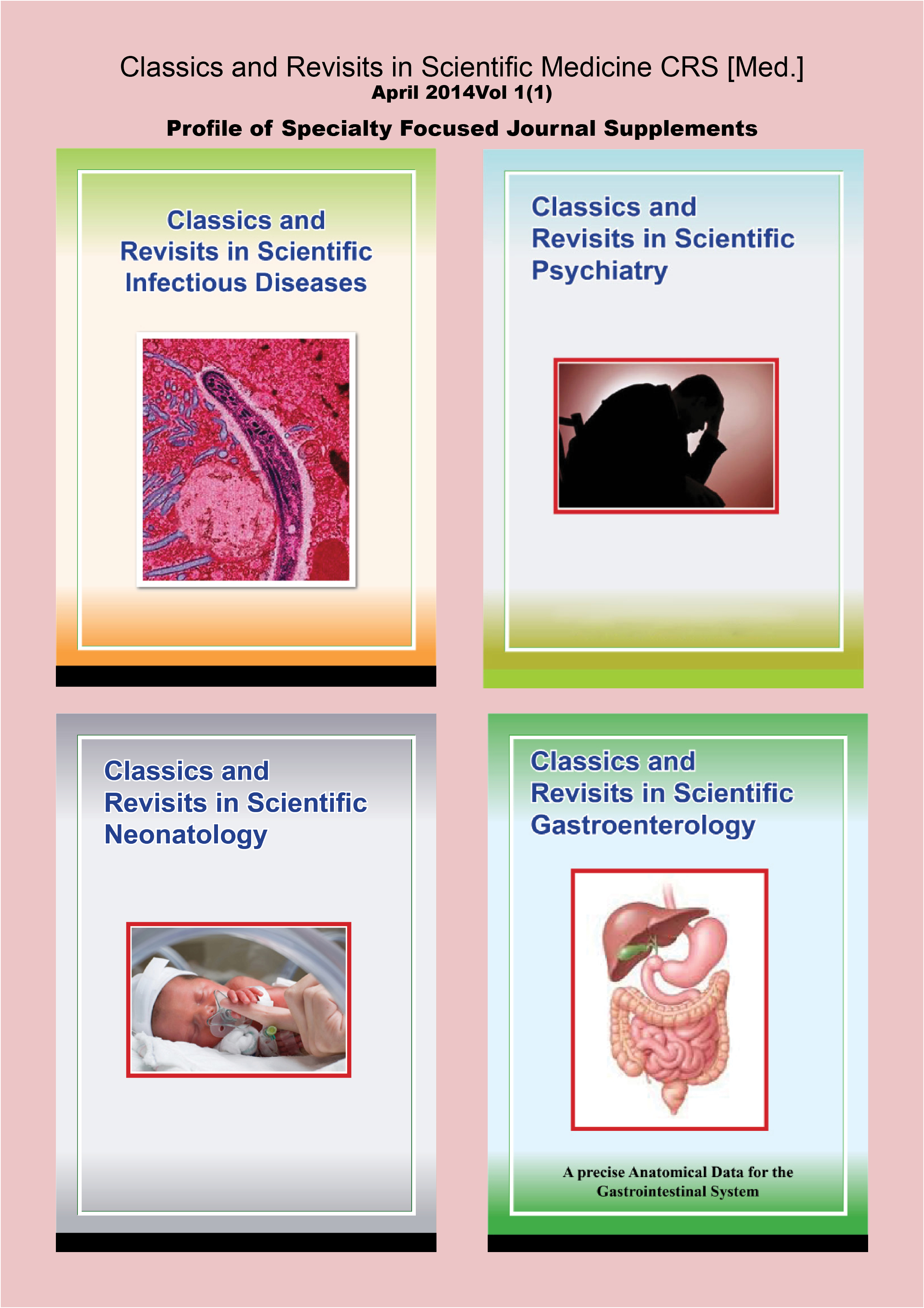
Classics and Revisits in Scientific Gastroenterology.CRS [Gast.Ent.]
A precise Anatomical Data for the Gastrointestinal System |
Profile of Specialty Focused Journal Supplements
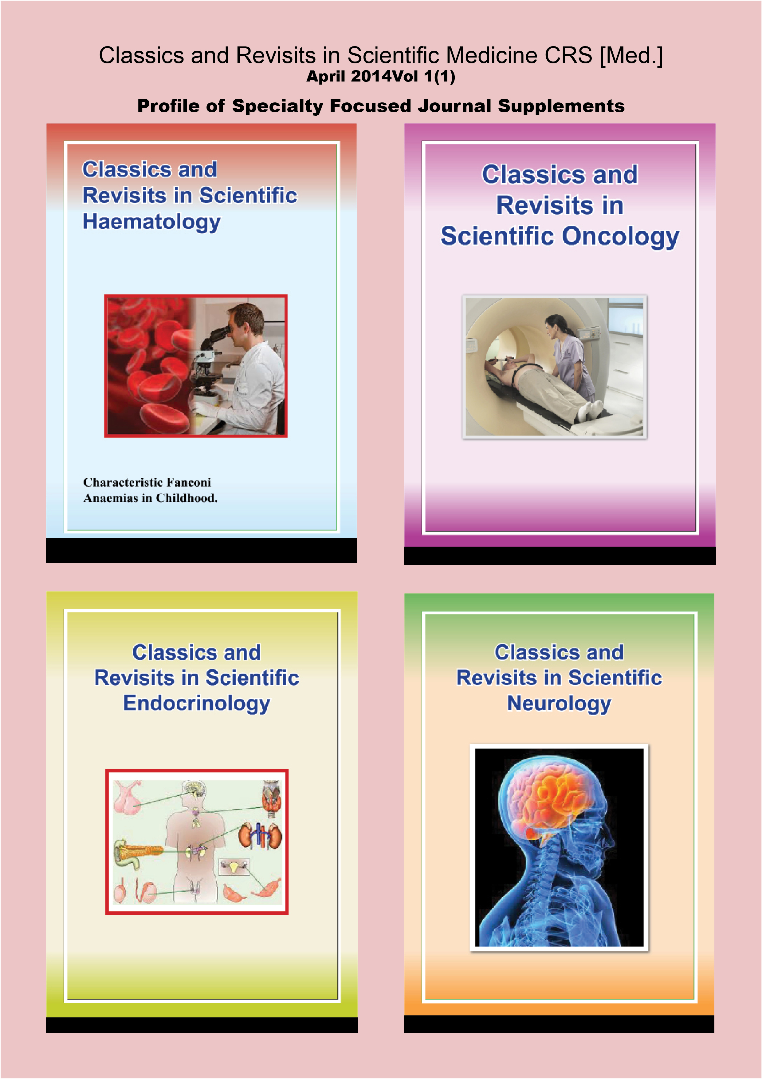
| Classics and Revisits in Scientific HaematologyCRS [Haem.]
Characteristic Fanconi Anaemias in Childhood.
|
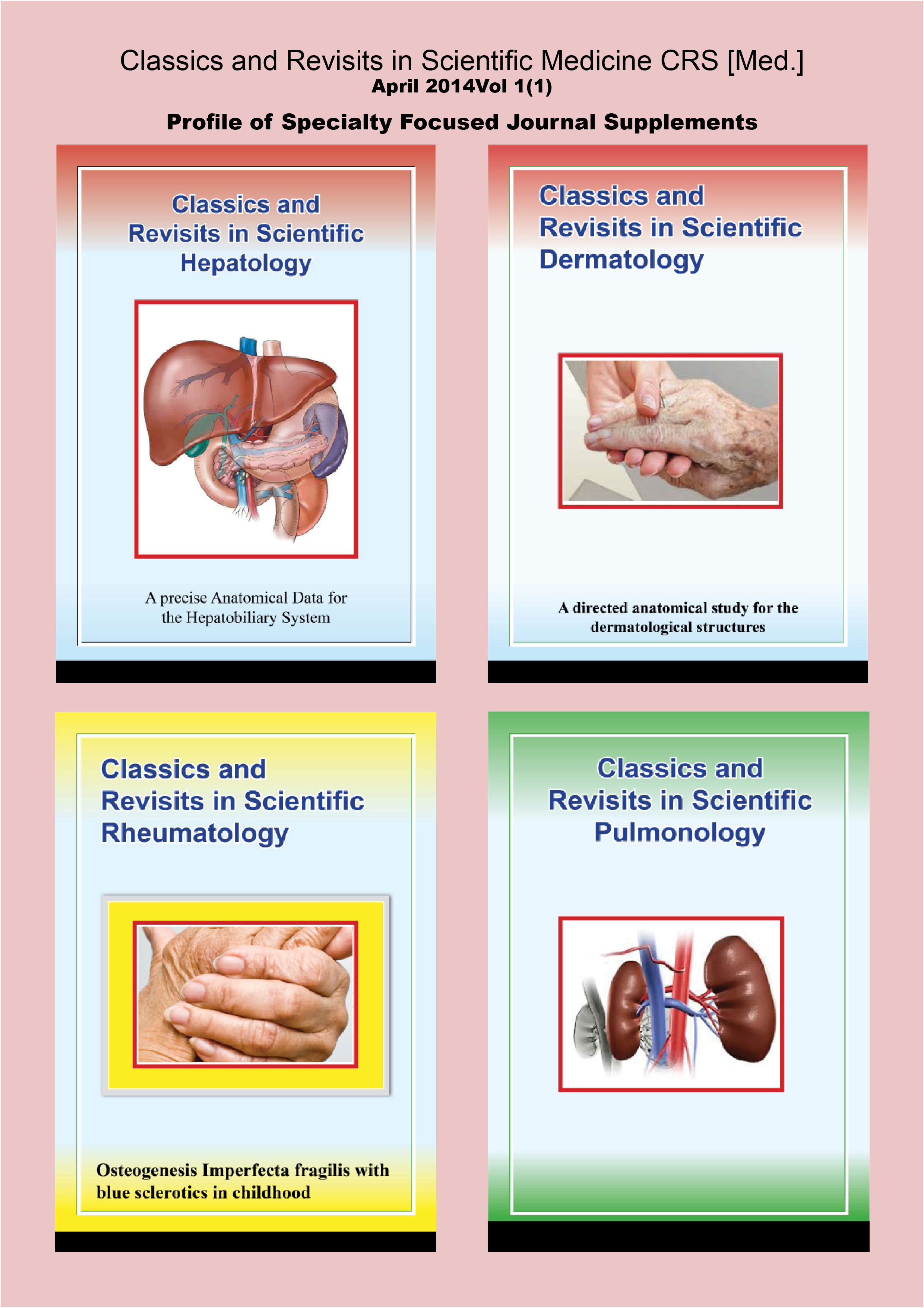
Classics and Revisits in Scientific Hepatology.CSR [Hep.]
A precise Anatomical Data for the Hepatobiliary System |
| Classics and Revisits in Scientific History of Medicine.CRS [His.Med.] | Classics and Revisits in Scientific Infectious Diseases.CRS [Inf.Dis.]
|
Classics and Revisits in Scientific Medicine CRS [Med.]
April 2014Vol 1(1)
Profile of Specialty Focused Journal Supplements
| Classics and Revisits in Scientific NeonatologyCRS [Neon.] | Classics and Revisits in Scientific NephrologyCRS [Neph.] |
| Classics and Revisits in Scientific NeurologyCRS[Neur.] | Classics and Revisits in Scientific Oncology CRS[Onc] |
| Classics and Revisits in Scientific Medicine CRS [Med.] April 2014Vol 1(1) |
| Profile of Specialty Focused Journal Supplements |
| Classics and Revisits in Scientific Preventive MedicineCRS[Prev.] | Classics and Revisits in Scientific Pulmonology CRS [Pulm.] |
| Classics and Revisits in Scientific Psychiatry CRS[Psych.] | Classics and Revisits in Scientific Radiology CRS[Rad.] |
| Classics and Revisits in Scientific RheumatologyCRS [Rheum.]
Osteogenesis Imperfecta fragilis with blue sclerotics in childhood |
Classics and Revisits in Interdisciplinary Medical Themes.CRS [Int.Disc.Med.Them.]
Proteus syndrome Cervical haemangiomas with intracerebral extensions |
Contents
Background and purpose of Journal and Supplements 2 .
List of Specialty Focused Journal Supplements Profiles 3. Journal and Supplements Editorial policy 4
Specialty Focused Journal Supplements Profiles. 7
Classics and Revisits in Scientific Cardiology Supplement (1) 14
Angina pectoris and Acute Chest Pain of Cardiac Origin.
Classics and Revisits in Scientific Rheumatology Supplement (1) 22
Osteogenesis Imperfecta Fragilis with Blue Sclerotics
in Childhood
Classics and Revisits in Scientific Interdisciplinary Medical Themes.
Proteus Syndrome in Childhood 29
Cervical Haemangiomas with Intracerebral extensions. 47
Classics and Revisits in Scientific Haematology. 49
Fanconi Anaemias in Childhood.
April 2014 1(1) Classics and Revisits in Scientific Medicine.
Back ground
Classics and Revisits in Scientific Medicine CRS [Med.] is new.
CRS [Med] is a balanced portal of continuing medical education information ,
Purpose
The intent is that CRSM [Med] could provide a fairly well effective reciprocal communicative pathway for continuing medical education development.
The text delivers the details of good basic and clinical medical practice clearly and concisely .It is a unique contribution to medical specialty practice development nationally, regionally and globally, in the context of ongoing health trends.Although the current directions in health care developments were highlighted, an unpretentious emphasis to quite often unavailable advanced technological innovations and models were reasonably. weighted.
Focus and Theme
Throughout the series efforts were made to balance the emphasis on the scientific fundamentals and the basic principles of management and prevention with research directions and recent advances in investigations and interventions.
In order to make it more reader friendly and handy, efforts were made to minimize the volume while at the same time not compromising the contents.
On this basis, suggestions were made that it should be a regular and frequent topical medical periodical in scientific basic and clinical medicine.
It is hoped that these compendia will be found most rewarding by medical practitioners, health professionals undergoing their undergraduate and postgraduate medical trainings and other health conscious professionals and individuals.
This is a legendary textbook of scientific medicine in a journal format.
Issues and Future Trends
In this edition of the CRS [Med], a preamble of the format of the publications is suggested.
However, it would be worthwhile to hasten to say that it is likely that there may be considerable alterations in these serial publications with regards to their academics structure, print layout, constituent parts, focus and certainly contents and periodicity.Therefore suggestions and opinions on contents inclusions and modifications are welcome.
In principle, publications would normally follow positive peer and editorial reviews, revisions and decisions.
Specialty Focused Journal Supplements
For clarity sake, the other associated serial medical scientific publications and channels of classics and revisits in scientific medicine, which will normally be published periodically as attached, merged or adhoc as isolated supplements to the parent journal CRS [Med] are alphabetically itemized according to the relevant medical specialties focus as follows:
April 2014 Vol 1(1) Classics and Revisits in Scientific Medicine
April 2014 1(1) Classics and Revisits in Scientific Medicine.
| Specialty focusedSupplements to JournalNumber | Specialty focusedSupplements to Journals Fulland abridged Title | Anticipated IssuesPer year/PublicationCycle | Specialty focusedJournalSupplementClassification |
| 01 | Classics and Revisits in Scientific Cardiology- CRS [Card.] | 12Monthly | Academic, clinical.professional&Scholarly |
| 02 | Classics and Revisits in Scientific Dermatology. CRS [Derm.]. | 12Monthly | Academic, clinical.professional&Scholarly |
| 03 | Classics and Revisits in Scientific Endocrinology. CRS [End.]. | 12Monthly | Academic, clinical.professional&Scholarly |
| 04 | Classics and Revisits in Scientific Hepatology . CRS [Hep.] | 12Monthly | Academic,clinical,professional&Scholarly |
| 05 | Classics and Revisits in Scientific History of Medicine. CRS [His.Med.] | 12Monthly | Academic, Informative.professional&Scholarly |
| 06 | Classics and Revisits in Scientific Infectious Diseases. CRS[Inf.Dis.] | 12Monthly | Academic, clinical.professional&Scholarly |
| 07 | Classics and Revisits in Scientific Interdisciplinary Medical Themes.CRS [Int.Disc.Med.Them.] | 12Monthly | Academic, Clinical, Informative.professional&Scholarly |
| 08 | Classics and Revisits in Scientific Neonatology. CRS [Neon.] | 12Monthly | Academic, clinical.professional&Scholarly |
| 09 | Classics and Revisits in Scientific Nephrology. CRS [Neph.] | 12Monthly | Academic, clinical.professional&Scholarly |
| 10 | Classics and Revisits in Scientific Neurology. CRS [Neur.] | 12Monthly | Academic, clinical.professional&Scholarly |
| 11 | Classics and Revisits in Scientific Oncology. CRS [Onc.] | 12Monthly | Academic, clinical.professional&Scholarly |
| 12 | Classics and Revisits in Scientific Preventive Medicine. CRS [Prev.Med,] | 12Monthly | Academic, clinical, Informativeprofessional&Scholarly |
| 13 | Classics and Revisits in Scientific Pulmonology. CRS [Pul.] | 12Monthly | Academic, clinical.professional&Scholarly |
| 14 | Classics and Revisits in Scientific Psychiatry. CRS [Psy.] | 12Monthly | Academic, clinical.professional&Scholarly |
| 15 | Classics and Revisits in Scientific Radiology. CRS [Rad.] | 12Monthly | Academic, clinical.professional&Scholarly |
| 16 | Classics and Revisits in Scientific Rheumatology. CRS[Rhem] | 12Monthly | Academic, clinical,professional&Scholarly |
| 17 | Grey Medical Literature. | Ad-hoc | Academic&Scholarly |
| 18 | News Letters,Brochures&Bulletins | Ad-hoc | Professional |
| 19 | Medical Health Updates | Ad-hoc. | Clinical &Professional |
| 20 | Other Miscellenous Publications | Ad-hoc | Clinical&Professional |
Classics and Revisits in Scientific Medicine. April 2014 1(1)
Editorial Policy
We are pleased to consider papers in the following categories.
Original Articles from primary and secondary research:
The original articles could normally be up to 3000 words.
The documentations of the original papers should follow the traditional IMRAD pattern introduction, methods/ patients, results.discussion.conclusion, acknowledgements and references
The contents are expected to be precise, factual and as concise as possible.
Illustrative colour, black and white photographs and line drawings could be used if they could make the contents more lucid.
The abstract or the summary section should be structured and limited to 250 words or less.
Review Articles:
Review articles would generally be less than 3000 words.
It should be well researched and referenced.
Short Reports and Brief Communications:
Short reports and brief communications may contain up to 1000 words.
No summary or abstracts are required, but authors could decide to include them.
The contents should be logically ordered, but division into sections is optional and up to ten references may be used.
This may be suitable for the presentation of descriptive studies, some personal experience/observation and some field studies.
Case Reports and Case Series
This is the format most suitable for the presentation of individual case reports of particular interest or an illustrative series of cases.
Correspondence to the Editor
This could be on any topic related to the subject areas of the journal, which could be new topics or based on articles which have appeared in the series.
Current Topical Issues, opinions, view points, annotations and perspectives
This part of the series will provide a forum for expert opinion on topical issues in the area relevant to the series.
Appropriate submissions may include proposals for action such as:
[I]-Sound clinical judgment and good practice.
[II]-Appraisal or critiques of ongoing practice or principles.
[III-Responses to published statements.
The length of submissions in this section should normally be less than 1000 words. The presentations are expected to be precise, succinct and factual.
Usually about 20 references or less will suffice.
Occasional papers
Manuscripts of reasonable academic or clinical relevance belonging to this category could be published in the journal or in its supplements.
April 2014 1(1) Classics and Revisits in Scientific Medicine.
Research Letters:
This is a good medium for communicating research findings, which are too brief for short reports or brief communications.
Supplements:
Supplements to the series by Consulting Guest Editors on emerging topical issues will be published with a specialty based focus, periodically and on ad-hoc basis.
Normally these specialty focused supplements will be published as attachments, merged or isolated and detached supplements to the parent journal CRS [Med]
Technical Notes, Methods and Devises:
Papers describing procedures, techniques or equipment adapted by authors to their own conditions of work are welcome.
Medical Highlights and Forum Digest:
Summaries or Abstracts of ongoing and emerging topical medical issues will be published in the relevant sections of the journal or its specialty supplements.
Normally, these abstracts and summaries will be limited to 350 words or less and are best presented unstructured.
Editorials:
Normally, each series will be accompanied by an editorial perspective which as much as possible will aim to illuminate on the most significant contributions in the series.
Periodically, and on ad hoc basis editorials by consulting academic clinical guest editors will also be included, in the journal series or its supplement.
Book updates, Book Reviews and the Academic Clinicians Book shelf:
The series will consider book reviews, book updates relevant to the specialty series. A book shelf highlighting other updated books relevant to the specialty could be mentioned or displayed.
Report of the Highlights of Meetings and Conference Proceedings:
Proceedings of meetings and conferences relevant to the theme of the series could be published along the main journal series or as a supplement.
The specialty relevant highlights of appropriate medical scientific meetings will be published in the journal series or as supplements.
Academic Clinicians Web-Blog watch and alert:
Clinicians and medical scientists could be made aware of interesting Web sites and web-blogs in their relevant specialties.
Expert Opinions and View Points:
This involves a best evidence based expert advise and answer on a difficult or ambiguous academic or clinical encounter in any area relevant to the scope of the series. Normally the question or the scenario would be presented in a contextual format.with no more than 250 words.
The interventional answer or the solution to the difficulty will then be printed along with the question in the journal or a supplement following consultations with academic clinical experts in the topic and specialty.
April 2014 1(1) Classics and Revisits in Scientific Medicine.
Ongoing and forthcoming medical conferences, courses and pathways to medical career opportunities and progressions.
The series will from time to time and on adhoc basis publish a list of accreditated medical conferences, courses and achievable pathways to medical career progressions and opportunities.
Health News
The series will from time to time publish a balanced scientific perspective of the medical health literature on emerging clinical issue of public health importance.
Profile of Medical Journals and other related Publications
Editors of Medical Journals or other relevant scientific medical publications could
proffer copies of their publications to the Classics and Revisits in Scientific Medicines.
A compilation of these publications would then be sent to our consulting guest editors, referees and reviewers for short listing, vetting and selection.The publications will then be showcased in the upcoming issues of CRS [Med] or its supplements.
Clinical debates and Round Table discussions.
Logical and constructive debates and discussions on emerging topical health issues, on clinical issues which could influence practice positively will be published in the journal or its supplements periodically or on adhoc basis.
Text Presentation Format:
Text should be double spaced and all pages should be numbered.
The first page should include the title and the full names of authors with their academic and professional qualifications and/or job title and institutional affiliations at the time of writing, postal and E-mail addresses, telephone and fax numbers or other relevant contact co-ordinates for correspondence.
Tables, figures and text boxes should appear on separate pages at the end of the document and be appropriately labeled.
Text Boxes
Text boxes should be used for tangential information, such as contact details or background information of organizations relevant to the article, lists of very short case studies or descriptions of related research/projects and details of elements of a study or project, such as a survey, which for reasons of clarity are best removed from the main body of the text.
References and Acknowledgements:
References and acknowledgements should each start on a new page.
References should be outlined using the Vancouver referencing style, as originally published by the International of Medical Journal Editors (ICMJE)
References should be listed numerically in the order in which they appear in the text. The numbers of the references in the text could be in superscript.
A corresponding numerical list of the references is supplied at the end of the text after conclusive remarks.
April 2014 1(1) Classics and Revisits in Scientific Medicine.
References should be adequate, but redundant references should be avoided.
The full list of references should include the names and initials of the authors up to six authors could be listed then followed by et al for the outstanding authors, title of the paper, journal title, year of publication, volume number, first and last page numbers.
References to books should give the book title, place of publication, publisher and year, those of multiple authorship should also include
chapter title, first and last page numbers and the names and initials of the editors.
Papers accepted but not yet published should be included in the references followed by (in press)
Those in preparation (and any submitted for publication), personal communications and unpublished observations should be referred to as such in the body of the text.
Consent:
[I]-Original sentences and paraphrases replicated and quoted from other authors should be cited in a standard manner and be referenced appropriately.
[II]-The consent of patients and approval of the protocol by an ethical
committee or relevant authority on ethical matters should be confirmed for human investigations.
[III]-Any potentially defamatory statement or those construed to be defamatory must be eschewed.
[IV]-Any tables or illustrations previously published should be accompanied by the written consent of the copyright holder to republication, an acknowledgement included in the caption and the full reference should be included in the list.
Patient confidentiality:
Where a patient might be identified through an illustration or from the text, it is essential that written permission is obtained from the patient and forwarded with the manuscript.
Proofs and off prints
Proofs are sent to authors designated to receive them and corrections should be made within specified guidelines.
Covering Letter:
A covering letter signed by all of the authors must be submitted with the articles, and original or secondary research papers.
E-mail submissions should be sent to the Classics and Revisits in Scientific Medicine editorial section with record of all approved authors e-mail accounts.
The letter must contain the following information:
[I]-Why the submission is appropriate for publication in Classics and Revisits in Scientific Medicine or its specialty focused supplements and what it adds to the existing body of medical scientific knowledge.
[II]-The manuscript category that the paper is for.
April 2014 1(1) Classics and Revisits in Scientific Medicine.
[III]-Confirmation that the paper meets the requirements for the category as laid out in this document, stating word count and confirming that references were formatted in the Vancouver style as detailed below.
[IV]-Confirmation that the paper has not been published elsewhere.
[V]-Declaration of competing interests or the absence of competing interests and the disclosure of all sources of funding.
[VI]-Original research must declare ethical approval from an appropriate body and consent from participants.
[VII]-Name the corresponding author and provide full contact details.
[VIII]-Authors should also include written consents from those individuals being acknowledged in their paper.
Key Words
Authors are advised to include about five main key words before their introduction, this will facilitate the indexing of the article.
Language Support
Ideally, authors are advised to seek the assistance of a native English linguist for a linguistic editorial revision, however if this is unachievable, the Editors will provide the necessary linguistic support.
Copyright.
Before publication, authors would be asked to transfer the copyright of their contribution to the publishers of the Journal or its relevant supplement.
Check Lists for Authors:
Please ensure inclusion of the following before the manuscript submission:
.Manuscript in Microsoft word.
.Manuscript corresponding authors name, qualifications, degrees and/or job title
contact co-ordinates outlining their institutional affiliations, postal address,e-mail and telephone/ fax numbers.
.Submission covering letter.
Consent forms where applicable.
Papers may be submitted in one of the following ways: :
.Two copies of the manuscripts typed and double spaced.
.On a disk accompanied by one printed copy.
.Via E-mail to:
Dr.Emmanuel.G.U. Onyekwelu
Honorary Academic Clinical Editor
Classics and Revisits in Scientific Medicine.
P.O.BOX 2696
Serrekunda Post Office
The Gambia.
West Africa.
Tel: +220/9908295/2207357804.
E-mail:euonyekwelu@hotmail.com
Website: cottageclinicdoctors.wordpress.com
April 2014 Vol 1(1) Classics and Revisits in Scientific Medicine
Journal and Supplements Subscriptions and off prints enquiries.
Subscription enquires and Editorial communications about prints and off prints of the Journal or its supplements should be addressed to the above address.
The concessional subscription rate for this issue is the equivalent of Ten Euros.
| CRS [Med]Classics and Revisits in Scientific Medicine.
|
| This novel edition of CRS[Med] is a composite compilation of continual medical educational resource consisting of a profile of four supplements to the journal with a medical specialty focus with a considerable weighting given to their editorial aspects and themes:Classics and Revisits in Scientific Cardiology.CRS[Card]Classics and Revisits in Scientific Rheumatology .CRS [Rhem.]Classics and Revisits in Scientific Interdisciplinary Medical Themes.CRS[Int.Disc.Med.Them]
Classics and Revisits in Scientific Haematology.CRS[Haem,] |
| The Classics and Revisits in Scientific Cardiology.CRS[Card] journal supplement Presents a Guest Editorial Perspective on Angina pectoris and Acute chest pain of Cardiac origin by the CRSM Academic Clinical Consulting Guest Editors. |
| Classics and Revisits in Scientific Rheumatology .CRS [Rhem.] Classics and Revisits in Scientific Rheumatology .CRS [Rhem.]With a paediatric slant dilates on the peculiarities of Osteogenesis Imperfecta fragilis with blue sclerotics in childhood as a Guest Editorial review article by the CRSM Academic Clinical Consulting Guest Editors.a paediatric slant dilates on the peculiarities of Osteogenesis Imperfecta in negroid Africans as an Editorial short communication. |
| Classics and Revisits in Scientific Interdisciplinary Medical Themes.CRS [Int.Disc.Med.Them] with a paediatric slant.This supplement includes a guest editorial review article on Proteus syndrome in childhood and cervical haemangiomas in children with an intracerebral extension as editorial review articles by the CRSM Academic Clinical Consulting Guest Editors. |
| In this Classics and Revisits in Scientific Haematology.CRS [Haem] supplement with a paediatric slant characteristic Fanconis anaemia in childhood was discussed as an Editorial review article by the CRSM Academic clinical consulting Guest Editors. |
| Specialty Focused Journal Supplements Profile April 2014Supplements to Classics and Revisits in Scientific Medicine.Volume No. 1 Issue No.1 |
|
|
Contents:
. Acute Chest pain of cardiac origin and Angina Pectoris.
. Osteogenesis Imperfecta fragilis with blue sclerotics in childhood.
. Proteus syndrome in childhood.
. Cervicalhaemangiomas with intracerebral extensions in childhood
. Characteristic Fanconi anaemias in childhood.
April 2014 Supplement 1 Classic and Revisits in Scientific Cardiology.
Angina Pectoris and Acute Chest pain of cardiac origin
Guest Editors: CRS [Med] Consulting Academic clinical Experts.
Definition:
Angina pectoris is an acute chest pain of cardiac origin due to a poor reception of oxygen supply by the heart muscles:
Aetio-Patho-physiology:
Angina pectoris usually ensues when the oxygen demands and requirements of the heart exceeds the capacity of the coronary arteries to supply an adequate amount of blood to the heart for the coronary cardiac perfusion.
In typical exertional angina pectoris, ischaemia results when myocardial oxygen demand is increased but supply is relatively compromised because of occlusion of more than fifty percent of the coronary arteries either directly through the obstruction by the eccentrically located arteriosclerotic plaque or following dynamic coronary artery vasoconstriction of the subjacent areteriosclerotically uninvolved vessel muscle wall.In the setting of atherosclerotic blood vessel pathology.
Usual Causes:
Artheriosclerosis, Coronary Artery Spasms, Anaemia and Erythrocytopaenia:
Coronary-Cardiac perfusion could be compromised or made ischaemic by a diminution in the caliber of the coronary arteries, erythrocytopaenia or anaemia.
The diminution in the caliber of the coronary arteries may be arteriosclerosis related or sympathetic dysautonomic resulting in coronary artery spasms.
The usual causes of erythrocytopaenia or anemia were reviewed elsewhere.
In intense anaemia or erythrocytopaenia, angina pectoris is likely because the number of red blood cells [which contains haemoglobin –the oxygen carrying molecule in the red blood cells or the amount of haemoglobin in themselves is below the normal range for the age and gender of the case leading to a diminution in the oxygen supply to the heart muscles and acute cardiac pain-angina. Other pathologies which cause an increase in the work of the heart and thus an augmentation of the oxygen requirements of the heart include:
Hypertension:
Intense to profound high blood pressure causes coronary artery vascular flow resistance following fibromuscular hyperplasia of the coronary artery wall and coronary artery fibromuscular spasms from reflex sympathetic dysautonomia.
Valvular Heart Disease:
Cardiac valvular stenosis or narrowing and valvular regurgitations or incompetencies especially of the aortic valves given its close anatomical relationship to the coronary arteries associated with rheumatic valvular heart disease or of other aetiological factors will lead to a net ineffective or insufficient aortic outflow and consequently coronary artery perfusion difficulties.
April 2014 Supplement 1 Classic and Revisits in Scientific Cardiology
Angina Pectoris and Acute chest pain of cardiac origin.
Cardiomyopathies:
Idiopathic heart muscles pathologies especially the hypertrophic obstructive cardiomyopathic forms leads to a thickening of the cardiac muscles -increasing the surface areas to be perfused, in addition to causing a compressive effect on the coronary arteries and a relative myocardial perfusion insufficiencies.
Arrythmogenic Right Ventricular Dysplasia or Type Four Cardiomyopathy could also cause arrhythmia related angina pectoris.
Hypercoagulable states:
The angina pectoris associated with hypercougulable states are usually a consequence of compromised ischaemic oxygenation of the heart muscles secondary to poor myocardial perfusion following sludging of poor blood flow due to its hyperviscocity.
Cardiac Dysarythmias:
Atrial and ventricular fibrillations are usually associated with angina pectoris because of the associated poor ventricular filling and hence cardiac output.
Aetiopathogenic Classification Scheme for Angina Pectoris:
Effort Angina Pectoris:
This is a chest pain or discomfort that typically occurs with activity or stress.
This pattern of angina is usually transient, but appears to be induced repeatedly by these activities or stress profile in a predictable and regular manner.
Postural Angina Pectoris
This is the form of angina that occurs on sudden recumbency due to the redistribution of the body fluid with an increase in left ventricular end diastolic pressure initiated increase in the cardiac work load and subsequent myocardial ischaemia.
Cardiac Syndrome X:
This form of angina pectoris usually affects middle aged menopausal women following hysterectomy and it is usually associated with ST segment depression on exercise thread mill test, but angiographically normal coronary arteries.
The prognosis for progression to coronary artery disease is the same as those for the normal population.
The cause of this pattern of angina is thought to be due to narrowing of the smaller coronary arteries or arterioles due to neurotransmitter or myocytes related chemical imbalance causing ventricular dysfunction or other forms of coronary arteriolar dysfunctions.
Hypersomniac Angina Pectoris:
This is angina related to sleep, especially the rapid eye movements [REM] sleep, it is probable that this form of angina is related to the relative hypoxia in this stage of sleep or the obstructive sleep apnea syndrome or associated nightmares.
April 2014 Supplement 1 Classics and Revisits in Scientific Cardiology
Variant or Prinzmetal Angina Pectoris:
This is an effortless Angina Pectoris and refers to episodic unpredictable angina which occurs during rest rather than during activities and it is usually accompanied by specific electrocardiographic changes such as transient ST segment elevation during the anginal attacks.
This form of angina is thought to be consequential to spasms of the large coronary arteries on the surface of the heart with or without atherosclerotic lesions.
Unstable Angina Pectoris:
This form of angina pectoris is metarmorphic.
It refers to angina pectoris with variable symptomatologies,given the constant symptomatology patterns of angina in any one individual, any augmentative change in its symptomatologies such as its severity, attack frequencies, uninduced events or whilst in the restive state should be taken seriously, and investigated, because the risk of a heart attack is usually considerable, because this may indicate a sudden narrowing due to a new cloth formation or rupture of an atheroma.
This form of Angina Pectoris is considered a form of Acute Coronary Syndrome.
Symptomatologies:
More often than not anginal pains are usually felt as a pressure or an ache beneath the breastbone-sternum.
This is usually interpreted as a chest discomfort or heaviness rather than pain.
The affected individual has an exertional chest discomfort assuaged by rest.
This discomfort may also occur in either shoulder or down either sides of the arm, the back, the throat, jaw or the teeth or as a burning sensation especially in the middle aged female.Anginal pains are usually transient, worse following cigarette smoking, emotional, exercise, exposures to cold or physical exertions especially after meals, walking in cold windy environment, sleep or nightmares
Patterns of anginal pains may be predictable or unpredictable.
Diagnosis:
Accurate historical details of the nature of the pain following the Ryles ten point algorithm questionnaire with its associated symptomatologies is very crucial for the exact diagnosis of angina pectoris. A physical examination in between events although mandatory may not be all that very useful. If an opportunistic examination is achievable during an event, a mild tachycardia, hypertension or dysarrthymia or specific or non specific ECG changes may be observable.
Coronary reserve is mostly compromised most in the subendocardium, ischaemia occurs initially in that region. If an ECG is undertaken during an episode of an anginal pain, ST segment depression suggestive of a subendocardial ischaemia will be observable.
April 2014 Supplement 1 Classic and Revisits in Scientific Cardiology
Angina Pectoris and Acute Chest Pain of Cardiac Origin.
Adverse prognostic features in Angina pectoris
The prognosis for angina pectoris is usually affected adversely by advancing age, extensive coronary artery disease, diabetes mellitus, hypertension, previous heart attacks, tobacco smoking, hypercholesterolaemia, events with excruciating chest pains, ventricular pump failures, the extent and severity of the coronary artery occlusions, abnormal ECG results, the prognosis for people with stable angina and efficient ventricular pump function, the prognosis is reasonably good.
Although the mortality rate for people with isolated angina pectoris is about 1.5%, it is considerably higher for those with the risk factors outlined above.
A good historical description of the symptoms may be enough to achieve the diagnosis of angina pectoris in most instances of the classically typically presenting cases.
Electrocardiogram
Electrocardiogram (ECG) will show ST segment changes as outlined above.
Interval ECGs are not usually very satisfactory, most useful information are usually achieved with the baseline and pharmacological exercise stress testing ECGs using a vasodilator.
Continuous ECG monitoring with a Holter monitor may detect abnormalities indicating symptomatic or silent ischaemia or prinzmetal angina which typically occurs at rest.
Exercise Stress Test.
An exercise stress test incites myocardial ischaemia, in patients with coronary artery diseases and offers some useful prognostic information to the clinician.
The patient who manifests ischaemia either clinically or electrocardiographically following the first few minutes of exercise is likely to have myocardial insufficiency related to coronary artery dysfunction or disturbances of its structural integrity.
An exercise stress test should be undertaken to define objectively the degree of exertion required to induce myocardial ischaemia due to coronary artery related haemodynamic disturbances. Patients with a positive exercise stress testing should be presumed to have a relatively considerable morbidity and should be referred for further angiographically gated imaging procedures.
Echocardiography.
In this procedure, ultrasonic waves could produce images of the heart called echocardiograms thereby outlining the size of the heart, the movement of the heart muscles, blood flow through the heart valves and the valvular functions during rest or related to exercise ischaemic conditions, Left ventricular functions is usually compromised by ischaemia
April 2014 Supplement 1 Classics and Revisits in Scientific Cardiology.
Angina Pectoris and Acute Chest Pain of Cardiac Origin.
Coronary Angiography:
For this investigation to be achieved, x-rays of the coronary arteries are taken after a radiopaque dye is injected.
Coronary angiography is probably the most accurate procedure for diagnosing coronary artery disease.
It is usually indicated when the diagnosis of angina pectoris or coronary artery disease is uncertain.
Coronary angiography is commonly used to help evaluate whether Coronary by-pass Graft or percutaneous coronary intervention is appropriate.
It could also be employed in the diagnosis of a spontaneous or drug induced coronary artery spasm.
Multimodal Computer Assisted Tomographic Scans.
Electron beam computer assisted tomographic scans could detect the amount of calcium deposits in the coronary arteries.
The amount of calcium present could be computed as the calcium score is roughly proportional to the likelihood of the person having an angina or a heart attack. The limitation of this procedure is that occasionally, calcium deposits may be present in people whose coronary arteries are not terribly compromised so the scores derived may not very much predict the need for a percutaneous coronary intervention or coronary artery bypass grafting.
Given the significant radiation exposure with this procedure, EBCT will not usually be applied universally, but would be restricted to those cases with a relatively significant high risk of death or acute coronary syndrome especially those with risk factors such as metabolic syndromes, diabetes mellitus, hypertension, hypercholesterolaemia, hyperuraceamia, abnormal, unclear or ambiguous test results.
Multidetector series Computer Assisted Tomographic scan
This is a new technique that employs a high velocity CT scanner comprising of multiple small detectors that could accurately identify coronary artery narrowing.
April 2014 Supplement 1 Classics and Revisits in Scientific Cardiology.
Angina Pectoris and Acute Chest Pain of Cardiac Origin
However the amount of radiation exposure is considerable.
It could also be used to ascertain the patency of a stent or a bypass graft, or to ascertain if atheromas have undergone dystrophic calcifications.
In addition it is very useful for displaying the cardiac and the coronary venous anatomy.
This procedure is absolutely contraindicated in pregnancy, or in patients who are unable to hold their breath for at least a quarter of a second or tachycardiac or cannot tolerate bradycardia or the drugs used to induce bradycardia.
If the test must necessarily be employed in patients with tachycardia, they may need to be allowed a cardio-selective beta blocker to regulate the heart rate below 70 beats per minute.
The technique is noninvasive and highly accurate in excluding coronary artery narrowing as a source of a person’s symptoms especially in those instances where the exercise stress test was unachievable or its results inconclusive.
Cardiac Magnetic Resonance Imaging
In cases of coronary artery disease, MRI could be employed to evaluate the narrowing of the coronary arteries, measure the blood flow in the coronary arteries and test how well the heart is being supplied with oxygen.
It could also be used to assess the abnormalities of the heart wall motion during exercise stress test which may signify a poor blood supply to that area.
MRI may also be used as a test of viability to assess whether areas of the heart muscles damaged following an acute coronary syndrome could recover.
It could be used to evaluate the heart and the large blood vessels arising from the heart such as the aorta and the pulmonary arteries.MRI is radiation free.
Management:
Treatment of angina pectoris depends partly on the stability and severity of the symptoms of the angina.
Management of Angina pectoris implies attempts to slow or reverse the progression of coronary artery disease by the rapid treatment and modifications of risk factors, such as hypertension, high cholesterol levels, cessation of cigarette smoking, obesity, recommendation of a low varied polyunsaturated fatty diet, which is low in highly refined sugar carbohydrates and exercise.
The patient will need to comprehend the pathological process to drive the motivation to modify coronary artery disease risk factors and to be able to know
where and when to seek medical assistance during aggravation of symptomatologies.
In stable angina with symptomatologies of mild to moderate severity, modifications of the above risk factors with occasional drug use may suffice.
If this approach is not enough to ameliorate the symptomatologies of angina considerably, a procedure to restore the myocardial perfusion by improving the coronary blood flow through a revascularization procedure should be recommended. When symptomatologies worsen rapidly, immediate hospitalization is usually required and the case should be evaluated for a acute coronary syndrome.
April 2014 Supplement 1 Classics and Revisits in Scientific Cardiology.
Angina pectoris and Acute chest pain of cardiac origin.
Pharmacotherapy:
Pharmacotherapy of stable angina pectoris is aimed to ameliorate myocardial perfusion and decrease the work load on the heart.
The pharmacotherapy of angina pectoris revolves around six groups of drugs.
A-The organic nitrates
B-The beta-blockers
C-The calcium channel blockers
D-The angiotensin-converting enzyme inhibitors.
E- The antiplatelet drugs.
F-Non-opoid and opoid analgesics.
The Beta Blockers
These groups of drugs antagonize the effects of the catecholamine hormones epinephrine and norepinephrine on the heart and other organs.
These hormones have chronotropic (increases the heart rate) and inotropic effects (increases the strength of the heart contractions, and causes the arterioles to constrict (there by causing the blood vessels to constrict)
The resting and the activity related augmentation of the heart rates and blood pressures are usually ameliorated by these groups of drugs, thereby the myocardial oxygen demand is reduced. This group of drugs reduces the risk of heart attacks and sudden deaths.The long term outcome for people with coronary artery disease is therefore improved.
[2]-Organic Nitrates
[A]-Short Acting Organic Nitrates
Nitroglycerin is a very short acting organic nitrate drug that dilates the blood vessels.Following the administration of this drug .the anginal events are usually ameliorated in about two to four minutes. It is usually quite handy and could be taken shortly in anticipation of an anginal event, however the effects of nitroglycerin usually lasts about 30 minutes. The dose could be administered sublingually (i.e. as a tablet placed under the tongue) or bucally (as a tablet placed next to the gum) or intranasally (as a spray inhaled through the nostrils)
[B]-Long Acting Organic Nitrates
Isorsorbide dinitrate is an example of a long acting organic nitrate
This drug could be taken as tablets orally about one to four times daily.
It could also be administered as nitrate skin patches and paste allowing cutaneous absorption over several hours.
However if they are taken regularly they may loose their ability to provide relief.
Usually a period of nocturnal drug holiday is usually appropriate for its long term anginal relieving effect to be maintained. The use of their effect is mainly aimed towards the relieve of anginal symptomatologies rather than prophylaxis against acute coronary syndromes or sudden deaths.
April 2014 Supplement 1 Classics and Revisits in Scientific Cardiology.
Angina Pectoris and Acute Chest pain of Cardiac origin.
[3]-Calcium Channel blockers
This group of drugs prevents the blood vessels from narrowing (constricting) and could attenuate coronary artery spasm and as a group have a hypotensive effect.
Calcium channel blockers are equally effective in Prinzemetal angina pectoris.
Verapamil and daltiezem has negative chronotropic and antiarrythmic effects.
This group of drugs on the average is used as third line drugs, if the above two groups of drugs are inadequate for symptomatic relief or are contraindicated.
[4]-Angiotensin Converting Enzymes Inhibitors
ACE inhibitors such as ramipril were often given to angina pectoris cases with the aim of attenuating the risk factors of acute coronary syndromes and sudden deaths rather than symptomatic relief of angina pectoris.
[5]-Antiplatelet Agents
Aspirin, disopyradimole, ticlopidine, and clopidegrel have antiplatelet effects and influence the function of platelets, so that they do not clump together and stick to the blood vessels walls.
Platelets which circulate in the blood, promote clot formation (thrombosis) when a blood vessel is injured.
If platelets collect on atheromas in an artery’s walls, the resulting clot could narrow or block the artery and result in an acute coronary syndrome.
Aspirin modifies the platelet function by reducing the tendency for its aggregation and thrombus formation thereby ameliorating the vascular occlusion associated with artheromatous thrombus formation.
Antiplatelet drugs are usually given to individuals with angina pectoris, unless there is a good reason not to, such as in individuals with bleeding diathesis or haematological dyscrasias of unclear significance or consequence.
The traditional teaching is for individuals with coronary artery syndrome to take cardio-aspirin 75mg daily in addition to clopidogrel daily to reduce the risk of an acute coronary syndrome. Another beneficial antiplatelet drug Ticlopidine, although with more frequent side effects could be employed in individuals who are allergic or intolerant to aspirin-clopidogrel combination.
[6]-Non-Opoid and Opoid Analgesics
Analgesia of varying strengths will invariably be a component of the anti-anginal pectorisarmamentarium.
Paracetamol, Aspirin, other non-steroidal anti-inflammatory agents and codeine or morphine could be employed in combination on individual cases as bridge therapy until appropriate beta blocker or calcium channel blocker is defined.
April 2014 Supplement 1 Classic and Revisits in Scientific Cardiology.
Angina Pectoris and Acute Chest pain of cardiac origin
In summary the objectives of the pharmacotherapy of angina pectoris is aimed at the reduction of myocardial oxygen demand during exercise or stress by the use of organic nitrates, beta blockers or calcium channel blockers
Promotion of maximal dilatation of the coronary arteries by the use of nitrates, calcium channel blockers.
Revascularization Procedures:
Percutaneous Coronary Intervention (PCI) angioplasty and Coronary Artery By Pass Grafting [CABG]
These procedures are the two main revascularization procedures employed in the management of angina pectoris where pharmacotherapy was unsuccessful.
However, it is worthwhile to note that these procedures are invasive anatomical techniques which aim to alleviate the immediate pathology, but they are not usually effective for the cessation of the progression of the underlying disease without influencing the risk factors favourably.
[A]-PCI is non-invasive and hence it is often preferred to CABG in a selected group of patients. It is particularly indicated in cases in one or two coronary artery vessel disease, especially when the blocked coronary vascular segments are not lengthy or extensive. However, increasing experience and more sophisticated techniques is allowing this procedure to be used confidently and more frequently.
[B]-CABG, is particularly beneficial to individuals with severe angina and coronary artery disease which were not amenable to relevant pharmacotherapy, good cardiac reserve and a normally functioning heart, no previous acute coronary syndromes, no chronic obstructive pulmonary disease, thrombophilias, carcinomatosis or other conditions that could preclude surgery or make it hazardous About nine of ten of such people will have a rapid and absolute relief of symptoms after surgery.
CABG could improve symptoms, exercise tolerance, and could decrease the number and dose of drugs needed
An electively planned CABG has an infinitesimal mortality at about less than 1 in 100 and a risk of asymptomatic or symptomatic intraoperative acute coronary event of about less than one in twenty.
Guest Editors: CRS [Med] Consulting Academic clinical Editorial Experts.
April 2014 Supplement 1 Classics and Revisits in Scientific Rheumatology.
Osteogenesis Inperfecta fragilis with blue sclerotics in childhood.
Guest Editors: CRS [Med] Consulting Academic clinical Editorial Experts.
An unusual preponderance of observational reports on children with phenotypic features consistent with Osteogenesis Imperfecta fragilis presenting covertly with a history of multiple long bone fractures and /or easy bruisability of the skin espoused an expert guest editorial consultations and review article on this theme.
Introduction/Historical Perspective
Osteogenesis Imperfecta or Vroliks syndrome is a heterogeneous group of inherited conditions arising from a variety of biochemical and morphological collagen defects.Willam Vrolik professor of Anatomy ,Pathological Anatomy and zoology at the Athenaeum Illustre(University of Amsterdam) discussed in his handbook of Pathological Anatomy(1842-1844) and Tabulae and Illustradam embryogenesis hominis et mamalium,naturalem tam abnorman (1844-49) a new born infant with numerous fractures and hydrocephalus.The Tabulae, had both Latin and Dutch texts, in the Latin text ,Vrolik used , the term Osteogenesis Imperefecta (in Dutch, gebrekkige beenwording) Vrolik also mentioned that the infant lived for three days and that both parents were suffering from Lues Universalis at the time of the baby’s birth .
On re-examination of the Index case.
The whole skeleton of the baby appeared poorly mineralized.
The family had large skulls, exhibited a broad and high forehead, large fontanels, frontal and temporal bossing, shallow orbits, and a protruding occiput.
At autopsy, the calvaria of the baby consisted of many wormian bones.
The tubular bones, although of normal length and only slightly curved, were very thin as were the ribs. All the skeletal structures showed one or more fractures and many fractures showed callus formation. Following further investigations on this topic, this case was retrospectively diagnosed as a case of Osteogenesis Imperfecta type II..
Willan Vrolik was one of the first scientists to realize that many skeletal dysplasias were not the result of a post natally acquired disease such as rickets or osteomalacia as many of his contemporaries believed. He thought that it might be due to insufficient intrinsic generative energy. He substantiated this by stating that in his study specimen a primary impairment of ossification is present and not a secondary degeneration.
The Index Case Description.
The descriptions given by Willem Vroliks in some of the specimens ,generated the term Osteogenesis Imperfecta and the eponym Vroliks syndrome for this genetic disorder characterized by increased fragility.[1]Fractures are frequent and its onset depends on the type of Osteogenesis Imperfecta.[2]Osteogenesis Imperfecta is a heritable disease that may result in bone fragility, increased joint laxity, decreased muscle tone, thinning of the skin, a bluish appearance of the sclera, scoliosis, deafness, dentigenosis imperfecta and other multiple abnormalities.
April 2014 Supplement 1 Classics and Revisits in Scientific Rheumatology
Osteogenesis Imperfecta Type 1
Osteogenesis Imperfecta type 1 is a disorder characterized by frequent bone fractures and deformities associated with fracture malunion and poor healing, blue sclera, normal or near normal stature and autosomal dominant inheritance pattern.
The incidence is thought to be one in about twenty thousand live births.
Males and females are affected equally Osteopaenia is associated with an increased rate of long bone fractures upon ambulation.
For some reasons not well understood, fracture frequency decreased dramatically at puberty and during young adult life but increases once again in late middle age.
Most observed patients presented with clinically overt symptomatologies prepubertally.
Progressive hearing loss, often beginning in the second and third decade, is a feature of this disease in about half the families.The proportion of Osteogenesis Imperfecta Type 1 patients with significant hearing loss rises steadily into middle age ,despite the general decline in fracture frequency .Conductive or mixed conductive and sensorineural hearing loss are more common in dominant pattern oseogenesis imperfecta than sensorineural hearing loss alone. Dentigenosis Imperfecta is observed in some subset of the patient population.
Mortality Figures.
Mortality could be significant even in hospitalized patients at about three percent..
Mortality figures are 3.7 times more in the negroid race.
OI related demises are more likely in women. Admissions at younger age connote more severe disease.
Children at a younger age are more severely affected and more difficult to manage.
They tend to fair better when referred to a dedicated paediatric unit [3]
A classification scheme and the inheritance pattern for OI
Inheritance pattern depends on the type. The classification of OI has been debatable.
The detailed aspects of its nomenclature have been reviewed.
Anecdotally there are four types of Osteogenesis Imperfecta, although with recent advances in molecular biological techniques, other sub-types have been deciphered with considerable over-lap between the subtypes. The mode of inheritance is autosomal dominant for Type1and VI and recessive for Type II and III. It has been reported in one of two twins. [4]Osteogenesis Imperfecta is a defect of Type 1 collagen which is present in many tissues including blood vessels.
The differentiation of child abuse and Osteogenesis Imperfecta is critical, because of medical and legal implications for the latter. [5, 6, 7]
Considerations for the Inheritance pattern in OI
The mode of inheritance of OI is variable and the clinical manifestations are heterogeneous. In addition to its bone involvement, osteogenesis imperfecta, may involve tendons, ligaments, skin scleras, dentin, fascia and blood vessels.
April 2014 Supplement 1 Classics and Revisits in Scientific Rheumatology
A modified classification scheme for OI
The most commonly used classification schemes are those developed by Sillence and Colleagues. [8]Type 1 Osteogenesis Imperfecta, which was previously called Osteogenesis Imperfecta tarda, is the most common form and is a relatively mild to moderately severe disorder , inherited as an autosomal dominant trait with varying penetrance and expressivity.
It is associated with blue scleras and hearing loss in about fifty percent of the cases [9]
Type 2 Osteogenesis Imperfecta is lethal in the perinatal period and was previously known as Osteogenesis Imperfecta congenita, it is the most severe form and it is inherited as an autosomal recessive trait.
Types 3 and 4 Osteogenesis Imperfecta have clinical manifestations of intermediate severity between that of types 1 and 2 and are often associated with bone deformities and varying degrees of short stature and hearing loss.
Hearing loss commonly occurs in patients with Type 3 OI, it also occurs in some patients with Type 4 Osteogenesis Imperfecta. [8, 9]Loss of hearing is one of the least constant major features of Osteogenesis Imperfecta, with occurrence that varies between twenty six and sixty percent[10]The association of Osteogenesis Imperfecta, hearing loss and blue sclera’s is often referred to as de Van der Hoene Kleyn syndrome, which was originally well described in 1918 by two authors. [11]This syndrome usually occurs in the late second and early third decades of life, younger patients have a mixed or sensorineural hearing impairment.[12]
Histopathological Features and Molecular genetic pathological aspects of OI.
Findings at histopathologic examination of specimens extracted from patients with Osteogenesis Imperfecta provide some clues and values to the Type of hearing Impairment that could be expected in these patients.Osteogenesis Imperfecta.
OI is one of the inherited connective tissue disorders that result in brittle bones.
The molecular defects reside in various mutations that lead to defective or decreased Type 1 collagen synthesis [9,13, 14]In greater than ninety percent of cases , Osteogenesis Imperfecta results from mutations in one of two genes,COL1A1 gene on chromosome 17 and COL1A2 gene on chromosome 7 which encodes the chains of Type I collagen.[15,16]
Clinico-pathological correlations.
Findings at the histopathological examination of specimens from patients with Osteogenesis Imperfecta provide clues to the types of hearing impairments that could be expected in these patients.Temporal bone specimens extracted from patients with osteogenesis Imperfecta congenita(Type II) demonstrated markedly delayed and deficient ossification in the three layers of the otic capsule.[17]Conductive hearing loss is expected in these patients and is mainly due to fractures that commonly involve the crus of the stapes or the handle of the malleus.These fractures lead to discontinuity of the ossicular chain or to fixation by ankylosis of the head of the malleus to the medial attic wall, Therefore the hearing loss in Osteogenesis Imperfecta could be of a conductive type and may not always be progressive[18] Adult onset hearing loss with Osteogenesis Imperfecta therefore could be of a conductive type and is not always progressive.
April 2014 Supplement 1 Classics and Revisits in Scientific Rheumatology.
Reports of Osteogenesis Imperfecta tarda (Type 1) are somewhat scanty since the availability of specimens depends on surgical procedures or autopsies.
The whole question on the f temporal bones morphologies related to deafness in OI was answered elsewhere. [19, 20, 21, 22]
Radiological Features of OI
Conventional radiographs of the tibia and fibular in IO could demonstrate multiple healed or healing fractures of the tibia , as well as non-united fracture deformities of the tibia or the fibular shaft .The bones are usually gracile and osteoporotic in appearance, which are some characteristics radiological features of osteogenesis imperfecta.
Further discussions on the collagenous defects in OI
The evaluation of children with multiple unexplained fractures is a clinically and radiological challenging one, because of issues concerning non-accidental injuries.
An inconsistent, non-correlating historical, clinical and radiological profile and cessation of further events will point more to a non-accidental injury as the cause of the pathological fracture. Abnormalities in collagen in other tissues other than the bones will account for clinical findings in addition to fractures, such as dentinogenesis imperfecta (fragile discoloured teeth), blue or grey colour of the sclera, increased joints laxities and a tendency to bruising(thought to be due to defective collagen of small blood vessels)
The distinctive features of OI
Reports on most of the observed cases, suggest that they probably belong to the Type I Osteogenesis Imperfecta or Type III.OI subsets.
The exact classification scheme could be accomplished, clarified and distinguished with exact certainty at molecular biological studies to determine if the collagen structure is qualitatively normal, but quantitatively reduced (Type I) or defective in (TypeIII) where it is abnormal. On epidemiological basis however, Type 1 OI would likely to be the diagnosis in the most of the observed reports because it is the most common and complicating fractures may not necessarily be present at birth.
Fracture patterns in OI
All types of fractures could be seen in osteogenesis imperfecta such as transverse, oblique and spiral fractures. Greenstick fractures and torus also occur commonly in children with mild to moderately severe defects..
Localizations to the lower limbs are particularly more common in osteogenesis imperfecta in comparison to normal children. Fractures in osteogenesis imperfecta could be metaphyseal.[23, 24] Rib fractures are also relatively common.
Of all these fracture patterns, metaphyseal fractures are those with the most implication for a multidisciplinary intervention, because of its strong connotations for
non-accidental injuries. [25]
The associated osteopathies in OI with their diagnostic significance.
Given that the primary defect in Osteogenesis Imperfecta is principally with the collagen.
The anecdotal evidence that osteopaenia, osteoporosis and diminished bone density are features in Osteogenesis Imperfecta could be unfounded. This is buttressed by the fact that precise densitometry and native densitometry in unfractured bones in Osteogenesis Imperfecta is generally within the normal reference range.[26]
April 2014 Supplement Classics and Revisits in Scientific Rheumatology
Osteopaenia and other abnormalities occasionally inferred, were usually multiple previous fractures and prolonged immobilization related. This diagnostic enigma is worsened by the fact that the assessment of bone density from X–ray radiological features is unduly unreliable [27] and as such it is usually not very useful overall in achieving the diagnosis of osteogenesis imperfecta especially in older children.
The diagnostic accuracy, reontenographic-densitometric correlations and precision is more distinct in infants and children with temporary brittle bone disease [28, 29] where they could assist to distinguish these conditions from Osteogenesis Imperfecta.
However lack of a standard reference range has compromised the applicability of densitometry with robust precision.
Wormian bones in OI
Other features that could espouse the diagnosis of Osteogenesis Imperfecta includes the presence of Wormian bones which are additional small bones within the sutures of the skull as was originally described in 1643 by Olaus Worm, a Danish anatomist.
In osteogenesis Imperfecta the number of Wormian bones are usually excessive and their distribution is in a crazy-paving pattern rather than a linear profile.
They are particularly most pronounced in the majority of cases with Type III Osteogenesis Imperfecta.
However Wormian bones could also be seen in excess in the milder types I, IV and other pathological states. The x-ray radiological techniques for the demonstration of Wormian bones and its implication for the diagnosis of Osteogenesis Imperfecta and other simulating conditions is an interesting topic.[30]The lateral and especially the Townes views are the most informative views in the assessment and demonstration of wormian bones and are particularly recommended as most appropriate for the exact assessment of Wormian bones ,especially in children with fractures, where the exclusion of a non-accidental injury will be expedient. The absence of Wormian bones will not be adequate to achieve an exclusion of Osteogenesis Imperfecta, especially in early infancy when it is notoriously absent as was confirmed by interval projections latter on.
Wormian bones are not notable features in temporary brittle bone disease.
Isolated periosteal reaction as a phenomenon is not all that a common feature of classical Osteogenesis Imperfecta except in relationship to fractures.
Occasionally, symmetrical metaphyseal sclerosis could follow fractures in Osteogenesis Imperfecta.
Other collagenous defects in OI with haemodynamic and functional consequence.
Being a collagen based defect, other features such as excessive joint laxity and cerebral aneurysms[31] and cardiovascular abnormalities especially pulmonary and aortic dilatation and regurgitation, mitral valve prolapse and subtle microscopic changes in the pulmonary artery and aorta could be associated features .These associations might not necessarily be present at birth, but usually they appear with the passage of time and plays a dominant role in the natural history of the disease process, especially with regards to deleterious consequences such as aortic dissections.[32, 33, 34] OI could be associated with bleeding diathesis which could be spontaneous or follow mild trauma. [35]OI could be associated with spondylolisthesis caused by extreme pedicle elongation.
April 2014 Supplement 1 Classics and Revisits in Scientific Rheumatology
The clinico-radiological distinction between OI and non-accidental injuries.
The Type 1 collagen is thought to make an important contribution to the structure and function of long bones.[36]From a Biomechanics perspective, bone is considered to be a non homogenous composite structure consisting at the very least of mineral and organic phases. In test of whole bone properties e.g. four point bending, the sum of the various phases of a composite structure usually contributes to the mechanical properties measured in other mechanical tests, one phase may predominate over the other.
Increased number of fractures with few or no bruises will point more to classical Osteogenesis Imperfecta rather than a non-accidental injury as the aetiological factor in cases of fractures, where as in non-accidental injury, bruising is much more common than fractures.[37, 38]
Although imaging is relevant to the diagnosis of Classical Osteogenesis Imperfecta it cannot be considered in isolation from the clinical findings, the historical events, the family history, the social assessment and the laboratory features.
Plausible pharmacological and physical therapeutic Interventions for OI
The beneficial therapeutic aspects of Osteogenesis imperfecta were contentiously discussed in the medical literature .Although no pharmacotherapeutic intervention has revolutionized the medical management of OI as such,however,the evidence for the beneficial effect of several pharmacotherapeutic interventions appears to be most convincing for the biophosphonates ,especially ,palmidronate or alendronate.
Also other reports have suggested the beneficial effect of the growth hormone in some selected group of children with OI, especially in aiding the affected children to attain an aesthetically acceptable linear growths, in other wise a subset that would have been disfiguringly stunted if the natural history of the pathology is uninterrupted with these pharmacological interventions.Orthopaedic and physical therapy in addition to modifications of functional, social professional and occupational ergonomics using assistive devices are other plausible interventional strategies.
Conclusion and Importance:
This editorial review article buttresses the need for clinicians to apply an in-depth evaluation, and scrutiny during patient’s encounter, to be able to decipher clinically covert pathological conditions and circumstances. Normally these evaluations would be most rewarding if a holistic multidisciplinary approach is introduced.
Guest Editors: CRS [Med] Consulting Academic Clinical Editorial Experts.
References:
[1.] Baijet B Aspects of the history of Osteogenesis Imperfecta (Vroliks Syndrome) An Anat 2002, 184(1).1-7.
[2.] Darmanis S, Bircher) Fractures of the acetabulum in Osteogenesis Imperfecta (Journal of Bone and Joint Surgery May 1.2006
[3.] Vitality,MG,Matsumoto ,Hiroko MA,Kessler ,Weinstein M,Hoffmann ,William BA,Roye,David P.Osteogenesis Imperfecta.Determining the demographics and the predictors of death from an inpatient population .Journal of Paediatrics orthopaedics.March 2007,27(2).228-232.
April2014 Supplement 1 Classics and Revisits in Scientific Rheumatology
[4.] O.Seriki.Osteogenesis Imperfecta in one of twins.Acta Paediatrica 21Jan, 59(3).340-342. and as a familial aggregation (Braga S, Passage E.Congenital Osteogenesis Imperfecta in three sibs (Human genetics) 1Oct1981, 58(4).441-443.
[5.] Klieman PK.Differentiation of Child abuse and Osteogenesis Imperfecta has medical and legal implications AJR (American Journal of Radiology May1990, 154. (1047-1048.
[6.] Bishop .N, Sprigg A, Dalton A, Unexplained fractures in infancy. Looking for fragile bones.Arch.Dis, Child, March 1, 2007, 92(3).251-256.
[7.] Cabral.W.A, Milgrom S, Leticha AD, Moriarty E, Marini JC.Biochemical screening of Type 1 collagen in Osteogenesis Imperfecta.detection of glycine substitutions in the amino end of the alpha chains requires supplementation by molecular analysis J.Med.Genet.August 1, 2006, 43.685-690.
[8.] Sillence D.O, Seen A, Danks DM, 1979, Genetic heterogeneity in Osteogenesis Imperfecta.J Med Genetm16.101-116.
[9.] Ablin DS, 1998, Osteogenesis Imperfecta, a review Can Assoc Radiol J.49.110-123.
[10.] Nager GT 1988, Osteogenesis Imperfecta of the temporal bone and its relationship to Otosclerosis.Ann Otol Rhinol Laryngol.97.585-593.
[11.] Van der Hoene J de Kleyn A.1918.Blaue Skleren, Knochen bruchigkeit Und Schwerhohrigkeit. Arch Ophthalmol 95.81-93. (German).
[12]Reidner ED, Levin LS, Holliday MJ.1980, Hearing patterns in dominant Osteogenesis Imperfecta.Arch Otolarngol.106.737-740
[13.] Byers PH, Steiner RD, 1992, Osteogenesis Imperfecta .Annu Rev Med 1992, 43.269-282
[14.]Kuivaniem H,Tromp G,Prockop DJ 1997,Mutations in fibrillar collagens(Type I,II,II and XI) ,fibril associated collagen Type IX and network forming collagen Type X cause a spectrum of diseases of the bone, cartilage ,and blood vessels. Hum Mutat 9.300-315
[15.] Byers PH, Steiner RD 1992MOsteogenesis Imperfecta Annu.Rev.Med.43.269-282
[16.]Skyes B,Ogilvie D,Wordsworth P,et al 1990,Consistent linkage of dominantly inherited Osteogenesis Imperfecta to the Type I collagen Loci.COL 1A1 and COL1A2,Am J Hum Genet 1990,46.293-307.
[17.] Berger G, Hawke M, Johnson A, Proops D.Histopathology of the temporal bone in Osteogenesis Imperfecta Congenita. a report of 5 cases. Laryngoscope 1985, 95.193-199)
[18.] Zajtchuk JT, Lindsay JR.Osteogenesis Imperfecta Congenita and tarda. A temporal bone report .Ann Otol.1975, 84.350-358
[19.] Sando I, Myers D, Havada T, Hinojosa R, Myers E Osteogenesis Imperfecta tarda and Otosclerosis, a temporal bone histopathology report. Ann otol 1981, 90.199-203
[20]Marion MS, Hinojosa R. Osteogenesis Imperfecta Am J Otolaryngol 1993, 14.137-138.
[21.]Tabor EK ,Curtin HD,Hirsh BE, May M .Osteogenesis imperfecta tarda.appearance of the temporal bones at Computed Tomography .Radiology 1990,175.1810183
[22.]Mafee MF,Valvasson GE,Ditch RL,et al .Use of the CT scan in the evaluation of Cochlear Otosclerosis.Radiology 1985,156.703-708
[23.] Hoddsmorth CE, Endahl GL, Soifer N, et al Comparative biochemical study of Otosclerosis and Osteogenesis Imperfecta .Arch oto-laryngol 1973, 98.336-339
April 2014 Supplement 1 Classics and Revisits in Scientific Rheumatology
[24.] Dent JA, Paterson CR.Fractures in early childhood.Osteogenesis imperfecta or child abuse? J Paediatr Orthop 1991; 11:184-186.
[25.] Ablin DS, Greenspan A, Reinhart M, Grix A, Differentiation of child abuse from Osteogenesis Imperfecta.Am J Roentgenol 1990.154:1035-1046
[26.] Paterson CR, Mole PA.Bone density in Osteogenesis Imperfecta may well be normal Postgrad Med J 1994:70:104-107.
[27.] Williamson MR, Boyd CM, Williamson SL Osteoporosis: Diagnosis by Plain chest film versus dual photon absorptiometry.Skeletal Radiol 1999; 19:27-30.
[28.] Miller ME. Temporary brittle bone disease: a true entity? Semin Perinatal 1992; 23:174-182.
[29.] Koo WWK.Sherman R, Succop P et al.Fractures and rickets in very low birth weight infants: Conservative management and outcome.J Paediatr Orthop 1989; 9:326-330.
[30.]Cremin B, Goodman H, Spranger J, Brighton P, Wormian bones in Osteogenesis Imperfecta and other disorders. Skeletal Radiol 1982; 8:35-38
[31.]Hawlik DM ,Nashesky MB,Ruptured cerebral artery aneurysm and Bacterial meningitis in a man with Osteogenesis Imperfecta.American Journal of Forensic Medicine and Pathology June 2006,27(2).117-120
[32.]Modestino G,Criscitiello M ,James A ,et al Cardiovascular abnormalities in Osteogenesis Imperfecta .Circulation 1965,31.255.
[33.]Wong R.S Follis.F, M Shively.B.K, Wernly .J.A.Osteogenesis Imperfecta and Cardiovascular diseases Ann.Thorac.Surg.November 1.1995, 60(5).1395-1397.
[34.] Moore.J.B, E.G.Wook and Kinkead.L.R.Ulnar artery aneurysm in Osteogenesis Imperfecta.J.Hand Surg Eur vol, February 1983.Original series, volume 15(1).91-95.
[35.]Ganesh.A, Jenny.C,Geyer.J.Shouldice M,Leven A, Retinal haemorrhages in type 1 ostegenesis imperfecta after minor trauma.Opthalmology.Volume 111,Issue 7.Pages 1428-1431.
[36.] Martin, R.B, Burr .D.B. (1989) The structure, function and adaptation of compact bone (Raven, New York), pp.57-84.
[37] McMahon P,Grossman W,Gaffney M.Stanitski C.Soft tissue injury as an indication of child abuse.J Bone Joint Surg 1995;77A:1179-1183.
[38.]Paterson CR, Monk EA.Long term follow up of children thought to have temporary brittle bone disease.Osteoporos Int 2000;11(Suppl 4):S47-S48
April 2014 Supplement 1 Classics and Revisits in Scientific Interdisciplinary Medical Themes CRS [Int.Disc.Med.Them.]
Proteus syndrome in childhood with a selective literature review.
Guest Editors: CRS [Med] Consulting Academic Clinical Editorial Experts.
The need for an Editorial review article on Proteus syndrome PS.
An editorial review article following academic clinical experts consultations on PS is undertaken on the basis of concerns evoked following observations on few compatible cases encountered over a considerable time. Given the progressive compressive, the vascular and malignant neoplastic implications of PS,a high index of suspicion should be expedient for an early and timely exclusion or diagnosis for PS,especially those cases presently covertly, or with minimal phenotypic expressivities or attenuations.
Case Definition
Proteus syndrome (PS) is a rare sporadically occurring hamartomatous disorder
associated with mosaic multifocal irregular asymmetric overgrowth of multiple body
tissues and cell lineages. The aetiology of Proteus syndrome is still idiopathic.
However, the predominant hypothesis is that PS is caused by a postzygotic mosaic
alteration in a gene that is lethal in its nonmosaic state.
Most malformations in Proteus syndrome have a predominantly ectodermal or
mesodermal origin.
Characteristic multifocal, multicentric, metachronous or synchronous overgrowths
(partial or regional gigantism, or hemi hypertrophy) in Proteus syndrome could involve
any body structure, especially the skin, bone, connective tissues, and fat.
Globally, published or unpublished reports of PS , are still very few.
A cluster of phenotypic features evoking the possibility of PS ..
On closer scrutiny,observations and follow ups, a couple of children presented with some of the spectral phenotypic dysmorphological features of PS such as lower limb hemi-hypertrophies, cerebriform cutaneous hyperplastic unilateral sole overgrowths, widespread hyperpigmented capillary haemangiomatosis on the limbs and extremities, saccrococcygeal, gluteal, perineal protuberant lipomatous masses,congenital absence of the gluteal cleft and progressive abdominal tumefactions.
Establishing PS as the most plausible diagnosis.
Following the observations of these features consistent with the recognized clinical diagnostic criteria for Proteus syndrome, the possibility of .this diagnosis was evoked and suggested, on follow ups ,with progressive enlargement of these structures the probability of Proteus syndrome was considered and discussed.
Rare embryonic neoplasms in PS
Following focused radiological imaging procedures the diagnosis of Proteus syndrome complicated by nephroblastoma, multifocal neuroblastomas and embryonic tumours was established in these cases.Although a range of malignancies have been previously reported in PS, embryomas were relatively uncommon especially Wilms tumour and Nephroblastom as they were demonstrated in the editorial observations
April 2014 Supplement 1 Classics and Revisits in Scientific Interdisciplinary Medical Themes.
The diagnostic and therapeutic difficulties in PS
The diagnosis and management of Proteus syndrome is challenging, enigmatic and
difficult and were controversially discussed in the medical literature.
Proteus syndrome could be associated with synchronous, metachronous,
multicentric, multifocal, phenotypic expressivities and intrabdominal embryomas
Literature search to aid the diagnosis. PS.
In establishing this diagnosis,the extant,and current medical literature were briefly reviewed to examine the rarity and strengths of these associated structural features in achieving the diagnosis of this syndrome against its other differentials. Other methodological issues concerning its epidemiology, presentations, diagnosis and management were also proffered and discussed.
Case definition.
Proteus syndrome (PS) is an extremely rare disorder of patchy or mosaic postnatal
growth dysregulation, primarily involving overgrowth. The disorder is thought to be
related to a somatic genetic alteration ,but its exact etiology is still largely unknown.
Proteus syndrome was named after the Greek god Proteus who could change his shape at will.This rare disorder is characterized by multiple lesions of the mesodermal tissues, fat cells,lymphatic and blood vessels (lipolymphohaemangiomas), overgrowth of one side of the body(hemihypertrophy), an abnormally large head (macrocephaly), partial gigantism of the feet, darkened spots or moles (nevi on the skin).
Historical Perspective and Nomenclature of interest.
Studies on Proteus syndrome commenced in earnest in 1978, when Bennett proposed a putative experimental model for the mode of inheritance in PS and other related syndromes associated by a dominantly determined genomically patterned inheritance with mosaicism sustaining the survival of a naturally lethal gene.[1]
Although some evidence of this syndrome was originally published in the medical
literature as early as in 1907 or before, the more modern medical description of the
disease was attributed to Cohen and Hayden, who identified the syndrome in 1979.[2]
To stress the polymorphic nature of the clinical manifestations of this new
syndrome, in 1983,Wiedermann coined the term Proteus syndrome after the Greek mythological god Proteus (meaning polymorphous), the Old man of the Sea who could change his shape at will to avoid capture. [3]
The Elephant Man had PS on retrospective analysis.
Anecdotally, Joseph Merrick, the Elephant Man described by Sir Fredrick Treves in 1884
celebrated by stage plays and movies with the same name, was initially thought to have
suffered from elephantiasis and latter on a diagnosis of neurofibromatosis was suggested
, but now believed retrospectively to have had a Proteus syndrome after a study of the preserved castings of his soles revealed a cerebriform cutaneous hyperplasia, a characteristic finding in Proteus syndrome. [4][5]It was in 1996 that the answer to what affected Merrick was determined with exact certainty . A radiologist, Amita Sharma, of the National Institutes of Health (U.S.A), examined very meticulously the x-rays and CT scans of Merrick’s skeleton preserved at the Royal London Hospital after his death.
April 2014 Supplement 1 Classics and Revisits in Scientific Interdisciplinary Medical Themes
Dr.Sharma in 1979. determined that Merrick had Proteus syndrome, an extremely rare disorder, she asserts that Merrick’s appearance, and especially his skeleton, portrays all the identified stigmata of the defect in their extreme.
Folk-lores on the Elephant man
Historical legends had it that the head of the elephant man was so large that his hat measured about three feet in diameter.
Undoubtedly, Joseph Carey Merrick the elephant man (1862-1890) wanted to be like other people. He often wished he could lie down and sleep, but because of the size and weight of his head he had to sleep sitting up. One morning in 1890 he was found lying down in bed on his back, dead. The weight of his head had compressed his airway, and he suffocated. He was aged 28 years old at the time of his death.
Retrospectively, in 1983, Weidemann suggested that the patient reported by Graetz in
1928 could have had Proteus syndrome. [6]and that another case reported by Temtamy and Rogers in 1976 had features consistent with PS [7]
An earnest Search for the phenotypic features compatible with PS
The same year, Weidemann et al described a new syndrome in four unrelated boys with a
constellation of clinical features of partial gigantism of the hands and or the feet, nevi,
hemihypertrophy, subcutaneous tumors, macrocephaly or other skull anomalies, possibly
accelerated growth and visceral affectations which were in keeping with Proteus syndrome. [3]Additional advancements in the comprehension of the
pathomorphology in Proteus syndrome led to relevant updates in 1984.[8]
That same year, Wiedemann and Burgio studied the extent and the nature of the dermal
changes in Proteus syndrome and asserts that they were papillomatous epidermal nevi piquantly aligned along the lines of Baloshka.[9]
Time lines of the Investigations on PS
Several Investigations were undertaken on PS from 1984 to 1990
Further investigations into the pathogenesis and phenotypic expressivity in
Proteus syndrome was undertaken by Lezama and Buyse et al still in 1984[10]
In the same year, Gorlin described another case with features consistent with Proteus
syndrome. [11]
Lipomatosis as an association of PS was highlighted In 1985
Mucke et al et al highlighted the dynamism of the clinical features of the Proteus syndrome by illuminating on the natural history of lipomatosis in I985 [12]The same year, further reports on the multi-focality of Proteus syndrome were made by Costa et al.Two cases were reported, with both having abdomino-pelvic lipomatosis.This was noted in one of the cases aged seven who retrospectively had a conjuctival dermoid at the age of thirty six months.Laparatomy at the age of seventy two months for an acute abdominal pain revealed a right iliac fossa lipomatosis and a twisted necrotic mesenteric fat as the implicated aetiopathogenic factor for the pain.Some morphological similarities of PS to the Bannayan-Zonana syndrome and linear sebaceous nevus syndromes were demonstrated. [13]With further case enrollments into these series in the same year by Costa et al, they asserted the paternal age independence of these cases in a study of ten cases, with a mean paternal age of 30(range 23 to 40) which were not unusually extreme.
April 2014 Supplement 1 Classics and Revisits in Interdisciplinay, Medical Themes.
The alignment of dermopathy of PS along the lines of Baloscka was noted in 1986
Happle pointed out that the lesions follow the dermal lesions along the lines
Baloschka.Happle had proposed that the likely putative aetiopathogenic pathway in PS is
an autosomal dominantly determined genomically transmitted lethal gene persisting by
mosaicism akin to hypothetically proven aetiopathogenic mechanisms propagating the
MCCune-Albright syndrome and probably the Schimmelpenning-Feuerstein-Mims
syndrome [14,15]
Association of PS with adherent lipomatosis and haemangiomas reiterated in 1987
Clark et al reported eleven cases of PS syndrome all with the remarkable
association of lipomatosis. [16] The same year in 1987, a case series was reported in Southern Africa by Viljoen et al [17], in the same year they mentioned that the surgical removal of lymphatic, fatty or hemangiomatous elements could be difficult ,usually resulting in unsightly scars and keloids,at about the same period a further case of a child aged fifty four months who was severely affected by Proteus syndrome was reported by Malamitsi-Puchner et al. [18] Between 1987 and 1988, further works were undertaken retrospectively by Cohen with regards to the Elephant man. [4, 19, 20]
Further aspects of the dermopathies of PS were highlighted in 1988,
Viljoen et al described the skin manifestations of the Proteus syndrome in all of their six patients, with marked hypertrophy of the skin of the soles, being a consistent and universal feature of this syndrome that the authors inferred that it was almost pathognomonic for the syndrome. In three cases, additional features such as large epidermal nevi and linear macular lesions with areas of depigmentations were also demonstrated.
On light microscopy, the affected skins from the soles were noted to have an elongation
of the cytoplasm of the basal cells.
Bannayan-Zonana syndromes suggested as a possible differentials of PS
The differential diagnosis of the Proteus and Bannayan-Zonana syndromes were discussed by Bailer et al in 1988In their examination of the relevant literature they noted a history of consanguinity in 5.56% of their cases with compatible Proteus syndrome.
The overlap within syndromes that include hamartoma as eloquent features intimates that
these syndromes may be aetio-pathologically related, probably with regards to
perturbations in the secretion of a hypothetical humoural growth factor or an irregular tissue or receptor responsiveness to this growth factor.[21]
Malignant transformation of PS lesions exemplified by progressions to testicular malignancy.
Samlaska et al. in 1989 reported an archetypical case and reviewed thirty four others reported sporadically. [22], Malamitsi-Puchner et al in 1990 gave an account of the
follow up of the cases that they studied and reported earlier in 1987.
A striking tissue overgrowth occurred after surgical interventions in this child who latter
developed testicular malignancy. [23]Still in 1990, Beluffi et al reported a case of Proteus syndrome with pelvic lipomatosis and illustrated the use of a computer assisted tomographic scan for demonstrating pelvic lipomatosis.
April 2014 Supplement 1 Classics and Revisits in Scientific InterdisciplinaryThemes The musculoskeletal associations of PS highlighted.
Further investigations in 1990 by Hotamisligil and Ertogan described the case in
a nine month old girl who in addition to other features, had soft tissue masses in the
paravertebral and gluteal areas with aggressive involvement of the spinal canal and a
hyperpigmented epidermal nevus with hyperkeratosis on one side of his body.
He had a bipedal and unilateral left palmar and dorsal macrodactyly with syndactyly of
the third and fourth left digits. In this instance, nephrogenic diabetes insipidus was
described in Proteus syndrome for the first time.[24 ]
Familial transmission of PS demonstrated.
Although traditionally considered a sporadic congenital disorder, some anecdotal reports
have proposed the possibility of a familial transmission in Proteus syndrome.
Goodship, et al in 1991 presented a possible case of father to son transmission of Proteus
syndrome. The son had a cranial hemihypertrophy, a lymphangioma, a lipomata, and
epidermal nevi, where as the father had a large lymphangioma resected from the right
side of his face as a child, given this association, the possibility that his father was a mosaic was proposed. [25]
The possibility of PS presenting with minimal phenotypic expressivity or attenuation highlighted.
Kruger et al in 1993, observed mild Proteus syndrome in a boy whose mother had very
attenuated manifestations of PS. She had facial asymmetry with hypertrophy of her right lower cheek and impaired facial expression in that region. He was not cognitively impaired, but he a had minimally expressed left unilateral superior labial hypertrophy and cheek, causing a perilesionally lateralized impaired focal facial expressivity, hypertrophy of the left arm, partial gigantism of the left middle finger, and a large subcutaneous swelling in the upper left abdomen thought clinically and sonographically to be a lipomata.Both this mother and her son had distinctive superiorly localized venous markings. [26]
The probability of spinal cord compression from related structural defects suggested in PS.
Also Skovby et al in 1993 reported two illustrative cases of the two plausible
pathways by which spinal cord compromise may develop in Proteus syndrome:
In the first instance, spinal stenosis resulted from an anomalous vertebral architectonics
such as angular kyphoscoliosis.In the second case, cord compression resulted from tumoural infiltration most likely froma paraspinal, intrathoracic angiolipomata.[27]
Somatic Mosaism and further associated organic structural defects in PS remarked.
Two unusual cases of PS were further reported by Cohen which supported the concept of
somatic mosiacism.In one case, a huge connective tissue nevus covered the anterior
thoraco-abdominal walls, in addition to multifocal calvaria hyperostoses.
In the other subject, linear verrucous epidermal nevi, epibulbar dermoids, and
hyperostoses were demonstrated. However no appendageal, digital or superficial plantar
enlargements were demonstrated in these instances.In the same year further review of selective aspects of Proteus syndrome, including uncommon neoplasms, pulmonary and renal abnormalities, brain malformations,and patterns of abnormal growth in the craniofacial skeleton were undertaken by Cohen.
April 2014 Supplement 1Classics and Revisits in Scientific Interdisciplinary Medical Themes,
At this point, the presence of external auditory meatal hyperostosis was taken as a very specific feature of Proteus syndrome. [28]
The multifocality of PS lesions demonstrated.
Smeets et al in 1994 reported another patient with a cranio-facial manifestation of
Proteus syndrome. In this case, multi-focal calvarial, facial bony, external auditory
meatal and mandibular hyperostoses were the principal regional features demonstrated.
Also a scleral tumor was demonstrated in this patient. These observations were interpreted as lending a further support to the hypothetical concept of somatic mosaicism. [29]
Further aspects of malignant transformations in PS were reiterated.further
Gordon et al in 1995, observed two patients with Proteus syndrome who progressed to
malignant transformations. One case had a probable mesothelioma, in addition to features
suggestive of thyroid papillary carcinoma, which was not ruled out with
exact certainty pre-mortem, this case latter died at the age of five whilst asleep, at
autopsy, a papillary neoplasm, most likely of mesothelial origin, involving the inferior
surface of the diaphragmatic musculature and infiltrating into the omentum, the pelvic
area, the scrotum and some of the mesenteric lymph nodes were demonstrated.
The other case had bilateral ovarian serous cystadenomas with nuclear atypia identified at six years three months of age, warranting.a right ovarian oophorectomy, invasion of the right fallopian tube was also noted. During this period a catalogue of uncommon neoplasms in Proteus syndrome was compiled and proffered. [30]
Multiple organomegalies and Visceromegalies as an association of PS.
Lacombe and Battin, in 1996 described two unrelated children diagnosed at birth with
isolated macrodactyly .At follow ups they were noted to have developed hemihypertrophy. Three dorsally located angiomas were seen in one of these cases,
a female child, when she was aged four years. The clinical features encountered in both cases were consistent with the specific diagnostic criteria for Proteus syndrome. [31]
Ceelen et al in 1997described a man with Proteus syndrome who sustained an enlarged
splenic rupture while horse riding. [32]
Splenomegaly as a common feature of PS
Biesecker et al in 1998 described their experience with eighteen cases with a
presumptive diagnosis of Proteus syndrome.Splenic hyperplasia was noted to be a recognized feature of PS. The spleen was enlarged in two of the eighteen cases, and another case with asymptomatic splenomegaly and thymic hyperplasia was known to them earlier on. However, they annotated that it was likely that hemi-hyperplasia commonly over simulates Proteus syndrome [33]
In the same year Biesecker et al described a distinctive subset characterized by static or
mildly progressive hemi-hyperplasia and multiple lipomata.
April 2014 Supplement 1 Classics and Revisits in Scientific Interdisciplinary Medical Themes.
A review of the diagnostic criteria for PS
The recommendations for the diagnostic criteria, differential diagnosis, and
guidelines for evaluation of patients with Proteus syndromes which were developed at a
workshop held at the National Institutes of Health in 1998 were reviewed by
Biesecker et al.The general criteria suggested as mandatory for the diagnosis of PS were mosaic distribution of lesions, a progressive course, and a sporadic occurrence.
Pathognomonic Features for the diagnosis of PS suggested.
Specific clinical manifestations also were suggested as necessary to meet the diagnostic
Criteria for PS. Connective tissue nevi, which were common manifestations in Proteus
syndrome, were considered almost pathognomonic for the syndrome, although they were
not always present in all cases, where the diagnosis of PS was made.
Other combinations of manifestations (e.g., epidermal nevus, disproportionate overgrowth, specific tumours were suggested to meet the diagnostic criteria. [34]
Describing other mutational variants of PS
Happle in 1999 suggested the designation elattoproteus syndrome for a disorder that he considered to be an inverse form of Proteus syndrome.
He described a seven year old boy with partial lipohypoplasia and patchy dermal hypoplasia involving large body surface areas. These areas of attenuated growth were similar to those described in many cases of Proteus syndrome. Paradoxically; however, he had a minimally expressed inordinate overgrowth defects.Happle proposed to explain this unusual phenotype by suggesting that there may be various allelic mutations in the putative gene locus responsible for Proteus syndrome, which gives rise to overgrowth of somatic tissues.
Pleioproteus and Ellattoproteus as mutational variants extremes of PS.
Such mutations could be called Pleioproteus alleles, a term derived from the Greek word pleion meaning plus. Conversely, the same gene locus may harbor alleles responsible for deficient growth of somatic tissues. Mutations in these points were referred to as Ellattoproteus alleles after the Greek word elatton, meaning minus.
Patients affected with Proteus syndrome may show classic overgrowth or a composite picture with Pleioproteus and Elattoproteus lesions or even an isolated putative previously uncharacterized elattoproteus phenotype [35]
Ophthalmopathy of PS highlighted.
De Becker in 2000 described the ocular manifestations in a case of Proteus
syndrome and reviewed the ocular features demonstrated in published cases. [36]
Also the same year ,Hodge et al described a ten year old boy with Proteus syndrome
who presented with an effusive pericardial disease, a panhypogammaglobinaemia, with a specific deficiency in IgG and IgA subsets accompanied by low levels of specific antibodies to pneumococcal and hemophilus type B polysaccarides, and global lymphopenia.Given that no other cause was found for this immune deficiency in this case of PS, the authors suggested that it may represent a previously unrecognized feature of Proteus syndrome. [37]
April 2014 Supplement 1 Classics and Revisits in Scientific Interdisciplinary Medical Themes
Cognitive impairments associated with neuronal migration disorders and ophthalmopathies demonstrated in PS
Further work on PS was undertaken by Gilbert-Barnes et al, in 2000, who reported an exceptional patient with Proteus syndrome in whom manifestations included multifocal meningiomas, polymicrogyria, and periventricular heterotopias, with both eyes having epibulbar cystic lesions. The retina showed diffuse disorganization with nodular gliosis, retinal pigmentary abnormalities, chronic papilledema, and optic atrophy. Other abnormalities noted included progressive cranial, mandibular, maxillary, and auditory canal hyperostosis, epidermal nevi, and mental sub-normality. His limbs, hands and feet were noted to be proportionate and otherwise normal. [38]
Deleterious Pulmonary embolism observed in PS
The same year, Slavotinek et al reported three patients with Proteus syndrome who died suddenly from pulmonary embolism related to different clinical circumstances.
The first patient, who was diagnosed with Proteus syndrome at the age of twelve years, had varicose veins, portal vein thrombosis, right iliac fossa vein occlusion and recurrent pulmonary embolism. At the age of twenty five years old he died from pulmonary embolism. The second patient was a nine year old male who collapsed and died at home. Autopsy asserted the cause of death to be pulmonary embolus associated with deep vein thrombosis. The third patient was a seventeen year-old female undergoing inpatient treatment for sinusitis when she suddenly died. Autopsy showed a large pulmonary embolus with no identified deep vein thrombosis.
The need for relevant historical enquiry for PS and evaluation for thrombogenicity to direct anti-thrombogenic prophylactic measures in PS reiterated.
Given the fatality related to these complications, Slavotinek et al promptly suggested that patients undergoing surgical procedures should be evaluated for coagulopathic potential to determine whether antithrombotic prophylaxis will be appropriate for them [39]Cohen in 2001 reviewed at least seventeen reported cases of premature deaths in Proteus syndrome and suggested that patients with this disorder and/or their families should make their health care providers aware of the risk of deep venous thrombosis and pulmonary embolism. [40 ]
Biesecker in the same year reported Proteus syndrome in a five year old case and made a selective literature review.[41 ]
Controversies on the role of PET Gene mutations in PS.
Further work on the molecular genetic aspect of PS was undertaken by Barker et
al in 2001, however they did not identify mutations in the PTEN gene in eight unrelated patients with classic Proteus syndrome, hence suggesting that the mutation exclusion hypothesis may not be unfounded.[42]
April 2014 Supplement 1 Classics and Revisits in Scientific Interdisciplinary Medical Themes
Obstipation demonstrated in PS, a consequence of infiltrative colonic lipomata.
Mackay et al in 2002 reported a twelve year old boy with Proteus syndrome who had presented with gross abdominal distension and severe intractable obstipation.
At an Axial T1- weighted MRI, a diffuse hyperintense tissue, signal typical of fat surrounding and separating bowel loops, with a lesional aspect extending posteriorly on the left into the paraspinal musculature, displacing the psoas muscle anteriorly was
demonstrated. At laparatomy a huge infiltrating lipomatous mass was identified encasing the left colon, including the rectum. [43]
Induced abortion with Misoprostol by a mother related to birth of a baby with PS
Mohameddbhai et al. also in 2002 reported the case of a newborn male Proteus
syndrome case whose mother had ingested misoprostol, an orally active prostaglandins,
at six weeks of gestation in an attempt to abort that conception. [44 ]
Further definitions of diagnostic criteria for PS
Turner et al in 2004 reviewed two hundred and five reported cases of Proteus syndrome.
Interestingly, of all these lot, only ninety seven were thought to have strictly met the diagnostic criteria of PS, eighty cases clearly did not meet the criteria; and although twenty eight cases had features suggestive of Proteus syndrome, there were insufficient clinical data to substantiate this diagnosis.
Follow up adverse events on putative PS cases using a stringent criterion.
On follow up, the reported cases that met the strict criteria for Proteus syndrome had a higher incidence of premature deaths and other complications (Scoliosis, megaspondyly, central nervous system abnormalities, tumors, otolaryngologic complications, pulmonary cystic malformations, dental and ophthalmic complications) compared to those in the non-Proteus group. Cases that met the criteria were more often males, which had implications regarding the hypothesis for the etiology and pathophysiology of Proteus syndrome.Cerebriform connective tissue nevi (skin lesions characterized by deep grooves and gyrations as seen on the surface of the brain, which are striking features when seen on the hands and feet) were considered characteristic.
Specific later onset tumours in PS
Specific tumors occurring before the second decade include ovarian cystadenomas
and parotid monomorphic adenomas. Lung cysts were also added as a criterion.[45]
The Controversies surrounding PTEN in PS clarified.
Thiffault et al in 2004 reiterated that the most plausible suggestion for the genetic
basis of Proteus syndrome is the Happle somatic mosaic hypothesis, although no somatic
mutations in candidate genes had been reported, because germ line mutations in the
PTEN gene had been identified in patients diagnosed with Proteus syndrome.
In the same year, Thiffault et al screened affected and unaffected tissue from six patients
with Proteus syndrome by direct sequencing of genomic DNA for germ line or somatic
mutations in the PTEN or GPC3genes. No intraexonic mutations were identified,
indicating that neither PTEN nor GPC3 was likely to have a major role in the etiology of
Proteus syndrome in this series of cases. [46 ]
April 2014 Classics and Revisits in Scientific Interdisplinary Medical Themes
Post zygotic mutations associated Mosaicism suggested as a causality factor in PS
Brockmann et al in 2008 reported a pair of monozygotic nine year old male twins
discordant for Proteus syndrome. The affected boy showed progressive postnatal
overgrowth of his right lower limb and foot with asymmetric progressive overgrowth of
single toes. There was a small cerebriform connective tissue nevus on his right fourth toe.
The phenotype was mild but still fulfilled the diagnostic criteria for Proteus syndrome.
The findings supported the hypothesis that this condition is caused by a post zygotic
mutation event resulting in mosaicism. [47]
Further discussions on establishing the diagnosis of PS:
Although the exact aetiology of Proteus syndrome is still idiopathic; however the
predominant hypothesis is that Proteus syndrome is a consequence of a post zygotic
mosaic alteration in a gene that is lethal in its non mosaic form, however, Proteus like syndromes were associated with PTEN mutations.
Somatic mutations and defective production or regulation of tissue growth factors or receptors as a causality factor in PS
The concept of a somatic mutation that affects the production or regulation of tissue
growth factors or their receptors could explain the sporadic occurrence, the random
distribution of overgrowth, and the wide range of findings within the phenotype.
Many features of Proteus syndrome overlap with other overgrowth syndromes.
A Proteus like syndrome was previously reported in association with a germ line and
tissue-specific somatic mutations in the PTEN [48, 49, 50] These PTENS genes are usually mutated in Cowden syndrome and in Bannayan-Riley-Ruvalcaba syndrome (BRRS)
Confounding diagnostic difficulties with PS due to other overlapping syndromes.
Some of the phenotypic features of this syndrome overlap with those of the other
overgrowth syndromes such as the Klippel-Trenaunay-Weber syndrome, the
Ollier disease and the Maffucci syndrome occasionally posing some diagnostic
challenges. [51]Although chromosomal alterations have not been demonstrated with routine cytogenetic studies as such in Proteus syndrome, however the karyotype in two patients with Proteus syndrome showed structural abnormalities of chromosome 16 and chromosome 1 in a mosaic distribution, further supporting the hypothesis of somatic mutation. This concept of a somatic mutation that involves tissue growth factors or their receptors could explain several aspects of Proteus syndrome, such as the mosaic distribution of lesions, its sporadic occurrence, the unaffected offspring from the affected individuals, and the existence of discordant twins. Despite the evidence implicating a somatic mutation, no causal gene mutations were known for Proteus syndrome.
However, ongoing advances in the comprehension of the aetiologic-phenotypic
correlations in the context of supposedly genetic defects appear to make the future more
certain. [52]
Striking a balance in the diagnostic criteria for PS.
The most appropriate criteria for the exact diagnosis of Proteus syndrome with much certainty were controversially discussed. Although under diagnosis could be more common, most stringent diagnostic criteria is commonly advocated over simplistic
overlapping generic ones to facilitate appropriate research investigations and utmost
clinical care.The strengths of most stringent criteria are that they define an entity with an
apposite level of congruity in clinical features and outcome which could lead to an
enhanced comprehension of the inherent putative pathogenic mechanisms in these cases [41,33,34 ]
Plausible diagnostic, prophylactic and therapeutic interventions for PS
The aggressive and disproportionate overgrowth in PS is progressive involving mostly the musculo-skeletal systems and these orthopaedic complications could be difficult and challenging to manage. An optimal intervention will imply an adequate knowledge of a spectrum of manifestations and complications of the disorder and a multidisciplinary approach involving the geneticist, surgeons and other specialists.
The diagnosis of Proteus syndrome could be achieved without routine cytogenetic
studies, which have demonstrated inconsistent chromosomal aberrations.
The determination of the molecular aetiology of this syndrome will be difficult, given its
extreme rarity, the abbreviated life span in affected subjects, and its sporadic occurrence,
making the conventional approach of traditional cloning less useful.
Novel molecular genetic approach for the diagnosis of PS.
The aetiology of this disorder could however be studied using various comparative molecular biological techniques such as c DNA arrays, genomic arrays, subtractive techniques, testing of candidate genes and other appropriate techniques.
In addition to testing for dysregulations of growth hormones and binding proteins in vivo.
A novel systematic diagnostic classification scheme for PS
An updated revised criteria was adopted for PS in 2014 [45], differing from that undertaken in 1999. [33] and other more generic less stringent ones which allowed the diagnosis of PS to be made in patients presenting ordinarily with hemi-hyperplasia. [34,53, 54].Its diagnosis requires the presence of all general criteria and various specific criteria, including the presence of the category A criterion or two category B criteria or three category C criteria. The affected subjects should have the following general criteria, mosaic distribution of the lesions, progressive course, and a sporadic occurrence.
The specific PS diagnostic criteria are as follows:
[I] +Category A-Cerebriform connective-tissue nevus.
[II] +Category B-Epidermal nevus; asymmetric, disproportionate overgrowth (limbs,
skull, external auditory meatus, vertebrae, and/or viscera); and specific tumours that
occur most commonly in the second decade (i.e., ovarian cystadenoma, parotid
Monomorphic adenoma)
[III]+Category C-Dysregulated adipose tissue (lipomas, regional lipohypoplasia)
, vascular malformations (capillary, venous, and/or lymphatic), lung cysts, and facial
phenotype..
The argument for a high index of suspicion for the diagnosis of PS
The variably mild form of this syndrome with a less eloquent phenotypic expressivity could be missed initially or entirely until catastrophic complication of DVT or PE renders it suspicious and the diagnosis now made in retrospect. Neuroimaging with Skull X-ray, CT or MRI will be most informative for those with significant facial dysmorphism, mental subnormality or epileptogenesis.Compressive neuropathies could follow tumourigenesis or bony exostosis.
April 2014 Supplement 1Classics and Revisits in Scientific Interdisciplinary Medical Themes
PS is a progressive and dynamic overgrowth condition.
Occasionally, the mild or moderate effects of Proteus syndrome, such as bening tumours, are present at birth. As the child grows and develops, the tissue overgrowth progresses and changes. This progression is often irregular, it is characterized by periods of major overgrowths and other periods of absent overgrowth.The effects therefore change over the course of a time.
PS is associated with ambulatory difficulties and compressive neuropathies.
However, most changes occur before adolescence, since tissue over-growths tend to plateau at that time.Patients with Proteus syndrome have difficulty ambulating because of toe macrodactyly, scoliosis, and joint instability, with frequent dislocations, expansive subcutaneous tumours and compressive neuropathies due to intraneural hamartomas.
Some patients may have persistent atelectasis, pneumonia, or symptoms of pulmonary
Strict diagnostic criteria, guidelines and algorithms has been proposed and accepted. Those features which are considered to have diagnostic values are considered because of the tremendous variability in the manifestations of Proteus syndrome.
These features could be classified as cutaneous or noncutaneous.
Cutaneous manifestations
Almost always, all patients with Proteus syndrome have at least one type of cutaneous
lesion, of which several different types have been reported. These skin lesions could
generally be classified into two groups, those that are congenital or neonatal in onset and are stable in the first group and those that are post neonatal and progressive in the second
group. In some patients, the presenting symptom may be one or more of the many types
of heterogeneous skin lesions that characterize Proteus syndrome.
These include cerebriform connective –tissue nevi, epidermal nevi, vascular hamartomas, lipomatas and hyperpigmented or hypopigmented areas. Epidermal nevi and vascular malformations are considered group one lesions, while lipomatas and cerebriform connective tissue nevi generally occur latter and are considered group two lesions.
Group 1 cutaneous lesions are characterized as follows:
Epidermal nevi are a common finding in patients with Proteus syndrome.
These manifest at birth as tan-to-brown, flat topped, hyperkeratotic or verrucous papules,
which run in a linear or whorled pattern along the lines of Blaschko.
Epidermal nevi are found asymmetrically, scattered around the body.
Histologic features are acantholysis and hyperkeratosis in a clinical lesion consistent with an epidermal nevus.
Congenital Vascular malformations are other common cutaneous features in PS.
These anomalies can be of venous, capillary, or lymphatic origin and include nevus
flammeus, angiokeratomas, cavernous hemangiomas, superficial and deep lymphangiomas, and varicosity of the superficial veins. [55, 56]
These lesions are developmental abnormalities and they grow proportionately through a
patient’s lifetime without regression .In an occasional patient these vascular malformations have been noted to expand beyond proportionate growth.
Prominence of veins may be enhanced in areas of regional lipodystrophy.
April 2014 Supplement 1 Classics and Revisits in Scientific Interdisciplinary Medical Themes.
Group Two cutaneous lesions are characterized by the presence of cerebriform nevi
Cerebriform connective-tissue nevus is one of the most common and characteristic
features of Proteus syndrome, although its presence is not required for diagnosis and its
presence alone is not pathognomonic for Proteus syndrome, although its presence is not
required for diagnosis and its presence alone is not pathognomonic for Proteus syndrome.
An occasional patient developed an isolated plantar cerebriform collegenoma who lacked the general diagnostic criteria for Proteus syndrome. These nevi have a relatively delayed onset, making the diagnosis of PS difficult in neonates and infants.
Cerebriform nevi occur as well-demarcated, skin coloured plaques with a cerebriform or
rugated appearance. Most often they occur in the cutaneous and subcutaneous tissues on
the palms or soles but have also been noted on the forearm, on the trunk, and inferior to the ala nasi.Lesions on the sole typically cause the most morbidity because they cause ambulation difficulties, are usually prone to ulceration and infection, and are malodorous.Histopathologically, the lesions consist of an irregular whorly non-lipomatous proliferation of highly collagenised cellular fibrous tissue.
Lipolymphohaemangiomas are characteristic features in PS
In PS adipose tissue abnormalities are common, resulting in lipomatas and areas of lipohypoplasia.Lipomatas occur as hamartomatous masses consisting of subcutaneous
tissue or a variable combination of adipose, lymphatic, and hemangiomatous components
(eg, lipolymphohaemagiomas) and are the second most frequent type of skin findings that
characterize Proteus syndrome. The soft subcutaneous tumours can affect any area of the
body but are most common on the head, abdomen, groin, or legs. They appear as soft,
skin coloured nodules or tumours.Patchy areas of lipohypoplasia or dermal hypoplasia
are also observed in some patients.Lipohypoplasia occurs as regions of skin with minimal
fat, while dermal hypoplasia appears depressed, red plaques in areas with prominent
Other non-lipomatous growths in PS
Cutaneous manifestations also include striking overgrowth anomalies, including
asymmetric hypertrophy of the face, part or all of one or both limbs (particularly
digits), the trunk, or any combination of these; often, hemihypertrophy of one side of the
body results. These abnormalities could also be the first to attract the parent’s attention
and cause considerable anxiety.Cafe au lait spots and areas of hypopigmentation or
hyperpigmentation with a linear or whorled arrangement are also hallmark skin findings
in persons with Proteus syndrome.Hypertrichosis and nail abnormalities are also seen.
Non-cutaneous manifestations in Proteus syndrome:
The most characteristic non-cutaneous findings in patients with Proteus syndrome involve skeletal overgrowth and include corporal hemihypertrophy, partial gigantism of the hands and or the feet, and skeletal anomalies such as long-bone overgrowth and scoliosis. Overgrowths of the hands, feet, or both were universal in one series of twenty four patients with Proteus syndrome. Arm or leg over growth is nearly always a constant feature of PS.
April 2014 Supplement 1 Classics and Revisits in Scientific Interdisplinary Medical Themes,
Cranio-facial abnormalities
These are fairly common and progressive and include cranial hemihyperplasia, hyperostosis of the skull or external auditory canal, craniosynostosis,
and unilateral condylar hyperplasia. Hyperostosis and unilateral condylar hyperplasia
were common, while cranial hemihyperplasia and craniosynostosis were rare.
A facial phenotype with dolichocephaly, long face, down-slanting palpebral fissures,
ptosis, low nasal bridge, wide or anteverted nares, and open mouth at rest has been
associated with mental retardation and seizures.
The other commonly associated neoplasms in Proteus syndrome
Specific tumours have been noted to occur with an increased frequency in patients with Proteus syndrome. In addition to being found subcutaneously lipomata could also infiltrate muscles and internal organs, including the heart, pancreas, spinal cord, and the pharynx. Other tumours noted to occur in several patients include ovarian cystadenomas, testicular tumours, and parotid adenomas. Other benign neoplasms, such as bronchial hamartomas and multiple meningiomas have been reported. Cystic lung disease is found in approximately 1:10 of patients with Proteus syndrome and could lead to a significant morbidity, with rapid progression, fatality could result. Renal cysts in PS have been previously described. Visceral overgrowth was reported previously, especially involving the spleen or the thymus. Internal vascular malformations of the gastrointestinal tracts, spleen, kidneys, and the testicles have been reported previously.Other occasional reports of intravesicular venous lesions associated with life threatening haematuria was previously proffered.
Other ophthalmopathies in PS
Many ocular manifestations were previously reported in patients with Proteus syndrome. The most commonly recorded of these ocular involvements included strabismus, nystagmus, and epibulbar tumours.Other ocular involvements include an over representation of a high degree of myopia, retinal pigmentary abnormalities, retinal detachment, cataracts, posterior segment hamartomas, retinal colobomas, heterochromia irides, and glaucoma.
Associated myopathies, cardiopathies and Immuno-deficiency states in PS
Other additional features in PS include asymmetric myopathies, congenital cardiac defects, malocclusions, hypodontias, and hypoplastic enamels. A congenital immunodeficiency of some sort was previously reported in a case of Proteus syndrome.
HHML is a differential of PS with much clinical consequence.
A very important differential diagnosis to be considered in these is hemihyperplasia
and multiple lipomatosis syndrome (HHML), because of the higher association with
nephroblastoma (Wilms tumours and possibly hepatoblastomas) in HHML.
The diagnosis of HHML with certainty will have implications for apposite screening of these patients for malignancies using standard screening protocols. [57]
April 2014 Supplement 1 Classics and Revisits in Scientific Interdisciplinary Medical Themes.
Early prophylactic surgical management are more appropriate interventions.in PS
The management of PS has been an inescapable but crucial medical dilemma,
especially with regards to the prolonged multiple staged orthopaedic interventions.
The rapidly progressive overgrowths are quite cosmetically disfiguring, unsightly and
probably mechanically and functionally incapacitating, seldomly or occasionally causing
compressive aerodynamically significant airway defects or neuropathies and rarely
malignant transformations. Despite several previous descriptions of early, intermediate or
delayed modified surgical interventions for these defects, overall short, medium or long
terms outcomes for these defects appear to be discouraging and unrewarding.
However, the evidence for a primordial intervention before the onset of catastrophic or
debilitating complications appears more convincing and satisfactory.
Interventions for PE in PS
The other factors of interest in the management of PS are the continual concerns with
deep vein thrombosis related thrombo-embolic phenomenology especially with regards to
its frequent occurrence peri or post operatively. The importance, strengths and the morbidity and mortality implications of these associations raised and buttressed the
argument for routine pre or perioperative anticoagulant prophylaxis and monitoring
of PS cases. However, this practice is more or less anecdotal and could occasionally be a
complex decision in the context of a sequence with a supposedly universal vascular
anomalies, real risk of bleeding especially in the association of Marriet Kaselbach
syndrome which is a potential complication of platelet consumption by the meshwork of
the vascular malformations.
Screening for Neoplastic conditions as plausible prophylactic strategy for PS
The other area worthy of adequate attention and consideration in the overall management of PS patients which were contentiously reviewed and addressed in the literature are those dealing with the most safe, cost effective and clinically useful way of detecting the tumourous events in PS early enough for appropriate interventions and outcomes to be most therauptically and prognostically rewarding. However, this decision is compounded by the fact that the list of plausible neoplasms which could complicate a case of PS are quite diverse and there appears to be no robust figures to suggest that earlier detection of these tumours or interventions by any means is actually rewarding.As such the time for active surveillance of these tumours should be weighed in favour of clinically and symptomatically suspected cases as against a routine procedure.
Genetic Counseling
Also issues concerning the genetic counseling of PS patients seem anecdotally based on
the mutational, nonmosaic lethality principle which infers a low recurrence risk for PS.
Finally, the physician investigators concerns of the most appropriate way of PS gene
mapping, identification and elaboration appears to be future challenges given its sporadic
Conclusions:
For a long time in the medical literature, issues concerning the exact diagnostic criteria of
PS in suspected cases have been topics of much debate, were controversially discussed,
inconclusive and not uniformly accepted. Although recently, these criteria were consensusly fairly well elaborated on, with some convincing recommendations derived. However, it is likely that these criteria will need to be modified from time to time as it has always been, to circumvent underdiagnosis or over diagnosis, the extensiveness and expressivity of the increasing number of overlapping syndromes make these diagnostic challenges likely, especially with further work in these areas or longer term follow up of putative PS probands and their families.
Relevance and Importance.
These editorial review discusses an unusual hitherto previously unaddressed co-existence and associations of embroyomas in PS.The diversity in its phenotypic expressivities and the variabilities in the chronology and temporality of their manifestations were reiterated.The rarity and strengths of its associated features as against its differentials especially hemihyperplasia and multiple lipomatosis syndrome (HHML) which is associated with embryomas were examined.[58] The other available literature on the other putative tentatively peculiar overlapping syndromes could be reviewed as a topic for debate in the near future.
Guest Editors: CRS [Med] Consulting Academic Clinical Editorial Experts.
References:
[1]-Bennett, D.: Rescue of a lethal T/t locus genotype by chimaerism with normal embryos. Nature 272:539 only, 1978.
[2]-Cohen Jr MM, Hayden PW: A newly recognized hamartomatous syndrome. Birth Defects Orig Artic Ser 1979; 15:291-296.
[3]-Widermann HR,Burgio GR,Aldenhoff P,Kunze J,Kaufmann HJ,Scirg E:The proteus syndrome. Partial gigantism of the hands and or feet, nevi, hemihypertrophy, subcutaneous tumours, macrocephaly or other skull anomalies and possible accelerated growth and visceral affectations.Eur J Pediatr 1983; 140:5-12.
[4]-Cohen Jr MM.The elephant man did not have neurofibromatosis.Proc Greenwood Genet Center 1987; 6:187-192.
[5]-Treves F: A case of congenital deformity.Proc Pathol Soc London 1885; 36:494-498
[6]-Graetz, I.Ueber einen Fall von sogenannter
totaler halbseitiger Korperhypertrophie.Z.Kinderheilk.45:381-403, 1928
[7]-Temtamy, S.A.; Rogers,J.G.: Macrodactyly, hemihypertrophy, and connective tissue nevi: report of a new syndrome and review of the literature.J.Pediat.89:924-927,1976.
[8]-Burgio, G.R.; Further and new details on the Proteus syndrome.Europ.J.Pediat.143:71-73, 1984.
[9]-Wiedemann HR, Burgio GR: Encephalocraniocutaneous lipomatosis and Proteus syndrome .Am J Med Genet 1986; 25:403-404.
[10]-Lezama, D.B.; Buyse, M.L.: The Proteus syndrome: the emergence of an entity.J.Clin.Dysmorph.2:10-13, 1984.
[11]-Gorlin, R.J ;Proteus syndrome J.Clin.Dysmorph.2:8-9, 1984.
[12]-Mucke, J.; Willegerodt, H.; Kunzel, R.; Brock, D.: Variability in the Proteus syndrome: report of an affected child with progressive lipomatosis.Europ.J.Pediatr.143:320-323, 1985.
[13]-Costa, T.; Fitch, N.; Azouz, E.M.: Proteus syndrome: report of two cases with pelvic lipomatosis.Pediatrics 76:984-989, 1985.
[14]-Happle R: Cutaneous manifestations of lethal genes. Hum Genet 1986; 72:280.
April 2014 Supplement 1Classics and Revisits in Scientific Interdisciplinary Themes
[15]-Happle R: Lethal genes surviving by mosaicism: a possible explanation for sporadic birth defects involving the skin.J Am Acad Dermatol 1987; 16:899-906.
[16]-Clark, R.D.;Donnai, D.; Rogers, J.; Cooper, J.; Baraitser M.: Proteus syndrome: an expanded phenotype.Am.J.Med.Genet.27:99-117,1987.
[17]-Viljoen, D.L.; Nelson, M.M.; de Jong, G.; Beighton, P.: Proteus syndrome in Southern Africa; natural history and clinical manifestations in six individuals.Am.J.Med.Genet.Genet.27:87-97,1987.
[18]-Malamitsi-Puchner, A.; Kitsiou, S.; Bartsocas, C.S.: Severe Proteus syndrome in an18-month-old boy.Am.J.Med.Genet.27:119-125,1987.
[19]-Cohen, M.M.Jr.: Understanding Proteus syndrome, unmasking the Elephant Man, and stemming elephant fever. Neurofibromatosis 1:260-280, 1988.
[20]-Cohen, M.M., Jr: Further diagnostic thoughts about the Elephant Man.Am.J.Genet.29:777-782, 1988:
(21]- Bailer, M.G.; Reidy,M.J; Wilson, W.G.Proteus syndrome versus Bannayan-Zonana syndrome: a problem in differential diagnosis .Europ.J.Pediat.148:122-125, 1988.
[22]-Samlaska,C.P.;Levin,S.W.;James,W.D.;Benson,P.M.;Walker,J.C.;Perlik,P.C.:
Proteus syndrome .Arch.Derm.125:1109-1114,1989.
[23]-Malamitsi-Puchner,A,;Dimitriadis,D.;Bartsocas,C.;Wiedemann,H.-R.:Proteus syndrome: course of a severe case.Am.J.Genet.35:283-285,1990.
[24]-Hotamisligil, G.S.; G.S.; Ertogan, F.: The Proteus syndrome: association with nephrogenic diabetes insipidus.Clin.Genet.38:139-144, 1990.
[25]-Goodship,J.;Redfearn,A.;Milligan,D.;Gardner-Medwin,D.;Burn,J.:Transmission of Proteus syndrome from father to son?Med.Genet.28:781-785.
(26)-Kruger, G.; Pelz, L.; Weidemann, H.-R.: Transmission of Proteus syndrome from mother to son? (Letter) Am.J.Med.Genet.45:117-118,1993.
[27]-Skovby,F.; Graham,J.M.; Sonne-Holm,S.; Cohen,M.M.,Jr.:
Compromise of the spinal canal in Proteus syndrome.Am.J.Med.Genet.47:656-659.
[28]-Cohen, M.M., Jr.: Proteus syndrome: clinical evidence for somatic mosaicism and selective review.Am.J.Med.Genet.47:645-652,1993.
[29]-Smeets, E.; Fryns, J.-P.; Cohen, M.M., Jr.Regional Proteus syndrome and somatic mosaicism Am.J.Med.Genet.51:29-31.
[30]-Gordon,P.L.;Wilroy,R.S.;Lasater,O.E.;Cohen,M.M.,Jr.:Neoplasms in Proteus syndrome.Am.J.Med.Genet.57:74-78,1995.
[31]-Lacombe,D,; Battin,J.: Isolated macrodactyly and Proteus syndrome. (Letter)Clin.Dysmorph.5:255-257.
(32]-Ceelen, W.; de Waele, J.; Kummen, M.; de Hemptinne, B: Non-operative management of a splenic laceration in a patient with Proteus syndrome.J.Accid.Emerg.Med.14:111-113,1997
[33]-Biesecker,L.G.;Happle,R.;Mulliken,J.B.;Weksberg,R.;Graham,J.M.,Jr.;Viljoen,D.L.;
Cohen, M.M., Jr.: Proteus syndrome: diagnostic criteria, differential diagnosis, and patient evaluation.Am.J.Med.Genet.84:389-395.
[34]-Biesecker, L.G.; Peters, K.F.;Darling, T.N.; Choyke, P.; Hill,S.; Schimke,N.;
April 2014 Supplement 1 Classics and Revisits in Scientific Interdisciplinary Medical Themes.
Cunningham,M,;Meltzer,P.;Cohen,M.M.,Jr.:Clinical differentiation between Proteus syndrome and hemihyperplasia:description of a distinct form of hemihyperplasia.Am.J.Med.Genet.79:311-318,1998.
[35]-Happle,R.: Elattoproteus syndrome: delineation of an inverse form of Proteus syndrome.Am.J.Med.Genet.84:25-28,1999.
[36]-De Becker,I.;Gajda,D.J.; Gilbert-Barness,E.;Cohen,M.M.,Jr.:Ocular manifestations in Proteus syndrome.Am.J.Med.Genet.92:350-352, 2000
[37]-Hodge,D,;Misbah,S.A.;Mueller,R.F.;Glass,E.J.;Chetcuti,P.A.J.:Proteus syndrome and immunodeficiency .Arch.Dis.Child.82:234-235,2000.
[38]-Gilbert-Barness, E.; Cohen, M.M.,Jr.;Optiz, J.M.: Multiple meningiomas, craniofacial hyperostosis and retinal abnormalities in Proteus syndrome.Am.J.Med.Genet.93:234-240.
[39]-Slavotinek, A.M.; Vacha, S.J.; Peters,K.F.; Biesecker,L.G.: Sudden death caused by pulmonary thromboembolism in Proteus syndrome.Clin.Genet.58:386-389,2000)
[40]-Cohen.M; Causes of premature death in Proteus syndrome (Editorial) Am.J.Med.Genet.101:1-3, 2001.
[41]-Biesecker,L.G.: The multifaceted challenges of Proteus syndrome.JAMA 285:2240-2243,2001.
[42]-Barker,K.;Martinez,A.;Wang,R.;Bevan,S.;Murday,V.;Shipley,J.;Houlston,R.
;Harper, J.: PTEN mutations are uncommon in Proteus syndrome.J.Med.Genet.38:480-481,2001.
[43]-Mackay, G.; Spitz, L.; McHugh,K.: Lipomatosis of the colon complicating Proteus syndrome.Arch.Dis.Child.86:265 only,2002.
[44]-Mohamedbhai,A.G.;Miyan,A.M.H.;Lacombe,D.:Neonatal Proteus syndrome?(Letter)Am.J.Med.Genet.112:228-230,2002.
[45]-Turner JT, Cohen Jr MM, Biesecker LG: Reassessment of the Proteus syndrome literature: application of diagnostic criteria to published cases. Am J Med Genet 2004; 130A:111-122.
[46]-Thiffault, I.; Schwartz, C.E.;Der Kaloustian, V.;Foulkes,W.D.: Mutation analysis of the tumor suppressor PTEN and the glypican 3 (GPC3) genes in patients diagnosed with Proteus syndrome.Am.J.Med.Genet.130A:123-127, 2004.
[47]-Brockmann, L.G.; Happle,R.;Oeffner,F;,Konig,A.: Monozygotic twins discordant for Proteus syndrome.Am.J.Med.Genet.146A:2122-2125,2008.
[48]-Smith,J.M;Kirk,E.P.E;Theodospoulos,G;Marshall,G.;Walker,J;Rogers,M;Field,M;
Brereton,J.J; Marsh, D.J.Germline mutation of the tumour suppressor PTEN in Proteus syndrome. (Letter)J.Med.Genet.39:937-940,2002.
[49]-Zhou,X;Hampel H;Thiele,H;Gorlin,R.J;Hennekam,R.C;Parisi,M;Winter R.M;Eng,C.:Association of germline mutation in the PTEN tumour suppressor gene and Proteus and Proteus-like syndromes. Lancet 358:210-211, 2001
[50]-Zhou, X.-P.;Marsh,D.J.;Hampel,H.;Mulliken,J.B,;Gimm,O.;Eng,C,:Germline and germline mosaic PTEN mutations associated with a Proteus-like syndrome of hemihypertrophy,lower limb asymmetry,arteriovenous malformations and lipomatosis.Hum.Molec.Gent.9:765-768,2000
[51]-Cohen JrMM, Neri G, Weksberg R: Klippel-Treaunay syndrome, Parkes Weber Syndrome, and Sturge-Weber syndrome: Overgrowth syndromes. New York: Oxford University Press, 2002, pp111-124.
[52]-Robin NH, Biesecker LG: Considerations for a multiaxis nomenclature system for medical genetics. Genet Med 2001; 3:290-293.
[53]-Darmstadt GL, Lane AT: Proteus syndrome.Pediatr Dermatol 1994; 11:222-226
[54]-Hotamisligil GS: Proteus syndrome and hamartoses with overgrowth.
Dysmorphol Clin Genet 1990; 4:87-107.
[55]- Tweed JV, Turner JT, Biesecker LG, Darling TN: Cutaneous manifestations of Proteus syndrome: correlations with general clinical severity. Arch Dermatol 2004; 140:947-954
[56]-Nguyen D,Turner JT,Olsen C,Biesecker LG,Darling TN:Evolution of skin lesions in Proteus syndrome.J. Am. Acad. Dermatol. 2005; 52:834-838.
[57]-De Baun MR, Brown M, Kessler L: Screening for Wilms tumor in children with high-risk congenital syndromes: considerations for an intervention trial. Med Pediatr Oncol 1996; 27:415-421.
April 2014 Supplement 1 Classics and Revisits in Scientific Interdisciplinary Medical Themes.
Cervical haemangiomas with intracerebral extensions in childhood.
Guest Editors: CRS [Med] Consulting Academic Clinical Editorial Experts.
Background and Purpose
The need for a multidisciplinary intervention in most cases in clinical medicine is further reiterated by another editorial annotation and perspective from the CRSM consulting guest editorial experts by cases of exophytic cervical haemangiomas with intracerebral extensions observed in some young children,
Editorial Annotation and perspective.
In this correspondence, unusual presentations of congenital Cervical Haemangioma with intracerebral corticomedullary extension, with acute respiratory distress, hypochromia and thrombocytopaenia in children is discussed. In such instances, early and timely interventions will have positive implications for guarding against occult Marriet-Kaselbach syndrome, high output cardio-respiratory decompensation, neurologically significant and multiple vital organic compressive-obstructive effects. In these circumstances, application of intralesional corticosteroids appears to have arrested the progressive course of these lesions, until interval extramural instrumentations were achievable.
Introduction
The descriptive epidemiology of Haemangiomas intimates that they are relatively common childhood tumours occurring in 1:10 to 1:20 infant’s .Female gender and extreme prematurity appears to be significant features of its epidemiologic associatiions.They are usually localized to the cutaneous or subcutaneous tissues. Although almost universally congenital, they are rarely fully developed at the time of birth. About 2 in 3 cases of haemangiomas are localized to the head and neck region. Most haemangiomas are present or appear during the neonatal period as soft rapidly growing solitary unifocal lesions or occasionally multifocal lesions.
Complications of Haemangiomas.
In haemangiomas rapid growth over six to nine months may occur to produce severe cosmetic disability, orificial obstructions, threatened viability of vital structures such as the eyes and multiple ulcerations. In less than 1: 10 cases, rapidly growing hemangiomas of the head and neck region could produce catastrophic complications, with aero-digestive tract obstructions especially following a sudden increase in size related to haemorrhages, causing visual, auditory, deglutinatory and vocal difficulties. Also the tumefactions could get secondarily infected through the ulceration of the overlying skins .If an arteriovenous malformative communications of a considerable capacity with a haemodynamic significance or consequence develops an intractable high output congestive cardiac failure could follow.
April 2014 Supplement 1 Classics and Revisits in Scientific Interdisciplinary Medical Themes.
Other deleterious consequences of Haemangiomas.
There are other several complications of great concern such as the Kasabach-Merrit syndrome. In this complication, an inordinate bleeding diathesis and purpura may result from platelet consumption, DIC, microangiopathic haemolytic anaemia.
Previously, internal organs have been reported to be occasionally involved, in
haemangiomas, however, intracerebral involvement in haemangiomas are rather an
unusual and rare phenomenon.
Thrombocytopenia, thrombocytosis, respiratory distress and occult bleeding diathesis from coagulapathy and thrombocytopaenia and severe recurrent iron deficiency anaemia appears to be real major concerns with significant lesions.
Associated naevus may initially be considered to be inconsequential but could occasionally pose cosmetic and aesthetic difficulties with the passage of time.
The natural history of haemangiomas and its simulating lymphogiomas
The natural history of haemangiomas peculiarly involves an initial proliferative growth
phase that lasts about half to one year and an involutional phase which is associated with
a slow regression of the haemagiomas, with about one half of the lesions resolving by sixty months and about 9/10th resolving by the age of ten years. A very important differential to consider in this case is a lymphangiomatous lymphatic malformations which could be unifocal or multifocal and commonly occurs in the facio-cervical region, axilla or thoracic, they are similar to haemangiomas in that they appear in infancy and childhood usually before thirty six months. However, they differ from them by the lack of spontaneous involution. They are equally obstructive and haemmorragic tendencies.
These lesions are quite extensive and diffuse making a stage evaluation for the exact
determination of the extent of the lesion and appropriate stage dissections imperative for
an utmost outcome.
Management
Intralesional steroids appeared to have arrested the progression of these lesions in some instances.In occasional circumstances however, management could be expectant, given that spontaneous resolutions has been known to occur. By the age of ten years 9 out of 10 cases would have resolved completely.
Systemic steroid applications would have been indicated otherwise if there were concerns with ocular integrity or ongoing concerns with airway obstructions, difficulties with feeding or defecation or in the Kasabach-Merrit syndrome
Although some recalcitrant cases may not be very steroid or radio therapeutically responsive. Administration of interferons appears to be very useful in catastrophic situations. Also Laser coagulations or sclerotherapy could be useful in haemangiomas but not usually in Lymphangiomas..Other agents like OTK 145 have been employed in selected cases with significant beneficial effects. In Kasabach-Merritt syndrome, treatment is supportive, platelets, cryoprecipitate and fresh frozen plasma, heparin and antifibrinolytic agents such as aminocaproic acid and traxenamic acid may be used.
In other instances, life threatening compressive effects such as severe respiratory distress or rising intracranial pressure with cardiopulmonary instability and intrabdominal hypertensive events has made urgent extramural instrumentations imperative.
Guest Editors: CRS [Med] Consulting Academic Clinical Editorial Experts.
April 2014 Supplement 1 Classics and Revisits in Scientific Haematology.
Fanconi anaemias in Childhood.
Guest Editors: CRS [Med] Consulting Academic Clinical Editorial Experts.
Background and Context:
Fanconi Anaemia-FA is a severe debilitating disease associated with a compromised life expectancy. Specific review on FA is crucial. This expert guest editorial review article by the CRSM academic clinical consulting editors is based on observational reports on a spectrum of compatible childhood symptomatic characteristic Fanconi akylating aplastic anaemias.
In the various subsets, overall aggregate annotations suggest that growth failures were universal, whereas dysmorphologies such as microcephaly, polydactyly, hypopigmented dermatoses were common associations.Rhizomelic short limbed dwarfism, hypodontias and caput quadratum were occasionally encounted defects. Bleeding diathesis, febrile neutropaenias, and pallor were predominantly featured phenomena.
Another notable feature of interest is that in individual cases, recurrent symptomatologies were invariable and somewhat predictable.
Multiple blood transfusions, antimicrobials, steroids. , testosterone, acyclovir and fluconazole were the most commonly applied interventions which lead to a partial remission in some cases.. Doubtful malignant transformations and acute catstrophic bone marrow failures were mutually exclusive events. Unprecedented demise was a notable phenomenon. Occasionally, recovery from FA could be spontaneous.
Case Definitions..
Fanconi aplastic anaemia is an inherited autosomal recessively transmitted, genomically determined instability syndrome, associated with multiple developmental structural and functional abnormalities, progressive bone marrow failure, leading to pancytopaenia and the requirement for haematopoietic bone marrow stem cell transplantation or increased morbidity or mortality as a result of defective haemopoesis and malignant predisposition to earlier onset acute myeloid leukaemias and latter onset solid stromal tumours. [1, 2]In Fanconi anaemia, cellular chromosomal instability, breakage and hypersensitivity to bifunctional DNA cross-linking agents especially mitomycin C is usually demonstrated, which usually forms one of the basis for its diagnostic criteria. [3]
Complementation somatic-cell hybridization genomic analysis in FA
For a long time, the relationship between congenital malformations, bone marrow
failures, cancers, chromosomal breakages and their derived complementation groups in
April 2014 Supplement 1 Classics and Revisits in Scientific Haematology
Fanconi anaemia syndromes were poorly understood. [4 ]
The principal pathophysiology in Fanconi anaemias, the most common of the familial congenital constitutional aplastic anaemias have been fairly well elucidated through major advances in complementation somatic-cell hybridization genomic analysis of FA cell lines which demonstrated and provided evidence for alphabetically designated complementation groups (FA-A, FA- B, FA-C, FA-D1, FA-D2, FA-E, FA-F, FA-G, FA-I, FA-J, FA-L) with implications for abnormal haemopoiesis and apoptosis in their mutant states especially for the FA-C [2] in addition to linkage to a specific pathognomonically genomic disease entity.
The distribution of the complementation subgroup subtypes.
The distribution of the complementation subgroup subtypes of FA is geographically distinct, in some regions the FA-A was the most frequently represented at 59%, where as in the other, FA-C was the predominantly featured subtype at 67%.
This distributional in homogeneity could have implications for mutation-screening
strategies in continental FA patients. [5]
The extensive genetic heterogeneity in FA and its influence on linkage analysis.
The implications of this extensive genetic heterogeneity for classical linkage analysis were further discussed in previous investigations. [6, 7, 8] however, a good part of its pathophysiology remains elusive.
Although it is a genetically inherited form of bone marrow failure syndrome, its presentation may not necessarily be congenital [9, 10], until the abnormal immune
responses implicated in its pathogenesis is triggered by genomic instability or by
environmental exposures to chemicals, drugs or viral infections or perhaps by
endogenously generated altered bone marrow cells, which then heralds its more florid
The molecular and geographic epidemiology of FA.
The pathobiology, molecular and geographical epidemiology with the relevant therapeutic options in Fanconi aplastic anaemia syndromes have been previously investigated, described, replicated and revisited in several settings [11,12,13,14] Previous occasional case studies and series suggested hospital and community based high incidence rates, however overall the disease prevalence was relatively low, such that longitudinal single city epidemiological studies were not logistically feasible, however more recent investigations achieved higher sample sizes but most of those were in adults.[15]Overall, FA is a relatively uncommon disease with an estimated prevalence of 1 to 5 per million and an approximated carrier frequency of 1 in 200 to 1 in 300 in most population studies [16] Although its diagnosis is probably underestimated, because its haematological features may be moderately expressed or they could present latter.
The influence of the Founder effect on the incidence of FA
Of growing interest in the elucidation of the drive behind the unusual preponderance of FA in some ethnic groups compared to others is the concept of founder effect associated with genetic and reproductive isolation. This phenomenon implies an increased risk of inheriting rare genetic disorders such as FA following the development of a high frequency of a mutant allele introduced into communities which are genetically and reproductively isolated and therefore consanguineously and endogamously married .
The carrier frequency in this subset was estimated to be 1 in 90 to 1 in 92[17,18]
Other previously described epitomes of the phenomenon of founder effect were demonstrated for a subset , where the carrier frequency was estimated to be about 1 in 77 [19,20]
Further recent investigations demonstrated the inordinately clustered overrepresentation
of the incidence of FA in the subjects of particular ancestry from heterogeneously
distinct descent, abode and ethnicity. However, the figures from those of subsets
with homogenous ancestries mostly of the complementation group A subset were
astonishingly extreme, probably the most globally at 1 in 64 to 1 in 70[21, 22]
Haplotype linkage disequilibrium as a causality factor in FA.
Other prominent clusters were demonstrated elsewhere, where the populace in
genetic isolation revealed clinical characteristics of the patients showing both intra-and
interfamilial heterogeneity suggestive of haplotype linkage disequilibrium for markers
flanking the FAA gene. This hypothesis may have implications for positional
cloning of this gene through haplotype disequilibrium mapping. [23]
The case for further investigations in FA
In most circumstances however, accurate estimates of childhood characteristic
Aplastic anaemia (Fanconi subset) Fanconi anaemia, its clinical profile, peculiarities, heterogeneity, complexities and the intricacies in its diagnosis and the feasible therapeutic options have not been previously analyzed, revisited or completely understood.
Reliable inference or reference review on Characteristic Fanconi akylating aplastic anaemia syndromes are crucial for interventional planning, hypothesis generation, research directions and focused hospital or population based interventions.
It could also offer some contribution to a better definition of its natural history. Furthermore, this annotation offers the opportunity to highlight and briefly discuss some of the commonly associated structural and functional anomalies in FA as was previously proposed [24] , given the relevance of a more lucid definition of these commonly associated anomalies in expediently advancing a rational prognostic scoring index which predicts the likelihood of progression to catastrophic bone marrow failures or malignant transformations as was determined previously[2,25,26] This editorial review article is a step to describe and characterize some of these aspects.
Case definitions, Observations and follow up of compatible FA cases.
On observation and follow ups, suspected cases with the diagnosis of compatible Characteristic Fanconi akylating aplastic anaemia syndromes would have achieved the clinical, radiological and pathological inclusion criteria of FA for descriptors, clinical features, predominant symptomatologies, laboratory features as was previously described by Camitta: [27]
With severe disease defined as the presence of two of three blood component counts
criteria of an absolute leukocyte count of<0.5×10 (9), platelet count of <20×10 (9) and a
reticulocyte count of < 1%.Extreme neutropenia (<0.2×10 9/L) defined very severe
aplastic anaemia, the other cases could be described as moderately severe aplastic
anaemia.The bone marrow biopsy had to be compatible with the diagnosis of aplastic
anaemia.Cases had to be analyzed for demographics, historical and medical information, especially with relevance to clinical features, recent and previous drug use and related infective episodes.
Clinico-Epidemiological diagnosis in FA
Further basic epidemiologic features of Fanconi aplastic anaemia bone marrow failure syndromes could be achieved by characterizing cases in terms of demographic variables, age, sex, medical history, clinically significant manifestation, proof of diagnosis, and prognostic criteria. Associated dysmorphologies were evaluated, studied, validated, analyzed and reported in a standardized manner using standard normograms from the dysmorphology databases.
Clinical features and associations of FA
Overall, presentations in FA were associated with the male factor, consanguinity, latter presentation,non-familial incidence,infective episodes, unusual cranio-facial features or inconsistent physical characteristics Bleeding diathesis especially with epistaxis was a common but less florid mode of presentation, where as massive haemoptysis.frank pulmonary haemorrhages and haematemesis,where less frequent, but clinically more alarming and deleterious.Lost cases were mostly related to massive haemorrhages or overwhelming infective events related to febrile neutropaenias.
Outcome of observed FA cases..
The presence of at least one associated anomalous dysmorphological defect is quite usual.Rhizomelic short limbed dwarfisms with delicate statute, microcephaly
pre-axial and post axial polydactyl, caput quadratum, absent radii,
hypodontias and abnormal gingival morphology were the most predominant dysmorphological phenomena. Abnormal pigmentation of the hair, the skin and the mucous membranes especially the lips were the most common intergumentary defect noted.
April 2014 Supplement 1 Classics and Revisits in Scientific Haematology
The diagnostic and the therapeutic aspects of FA.
For the suspicion of Characteristic Fanconi anaemia to be evoked, the blood film and bone marrow morphologies has to be compatible with that of bone marrow failure syndromes.
Undertaking radiological evaluations would be expedient to exclude respiratory tract infections or complicated organizing pneumonias or features suggestive of pulmonary infarctions or haemorrhage, it may equally exclude or suggest erythrophagocytic lympho-histiocytosis-X rather than FA as the plausible diagnosis to be pursued.
X-ray radiological surveys could be worthwhile from the context of suggesting absent radii. Or other more covert musculoskeletal defects.
Transthoracic and abdominal ultrasonic echographies or tomograms could be useful to exclude the possibility of other linked congenital visceral abnormalities.
Routine cultures and biochemical studies would have positive prophylactic, therapeutic and prognostic implications for those with persistent febrile episodes despite applications of broad spectrum and specific antimicrobial. Most patients would be hospitalized at a point, and most of the hospitalizations will be associated with whole blood transfusions, and most cases could be managed conservatively at the first instance.
The historical perspective of FA.
Fanconi aplastic anaemia syndrome, a form of Constitutional aplastic anaemia was
earlier described by Erlich towards the end of the 19th century. [28]
Constitutional aplastic anaemia could be associated with congenital dysmorphology
(Fanconi subset) or it may not be associated with it. In 1919 Smith reported aplastic
anaemia in a six year old boy associated with marked nuchal, genital, umbilical and areolar skin pigmentation. [29] Most cases of constitutional aplastic anaemias were usually autosomal-recessively transmitted, although an occasional case report suggested the possibility of an autosomal familial dominant inheritance pattern [30]
Fanconi in 1927 described three brothers aged five, six and seven with hyperchromic
anaemia, leucopaenia, thrombocytopaenia, haemmorragic diathesis, microcephaly, testicular hypoplasia, convergent strabismus, exaggerated deep tendon reflexes and generalized brown, melanin like pigmentation of the skin but not of the buccal mucosa. [31]
The Haemopoetic stem cell line defect in FA and its therapeutic implications.
Fanconi anaemia bone marrow syndromes usually arise as a result of damage to precursor cells so that they cannot supply an adequate quantity of haemopoetic cells .This damage could also be at the bone marrow stromal level (i.e.the bone marrow microenvironment) which is now regarded to be equally important in the pathogenesis of aplastic anaemia.The spectrum of clinical and laboratory features are implicit on the stage of the haemopoiesis where the maturational sequence was disrupted. Earlier damage will result in uniform pancytopaenia, whereas latter disruptions after haemopoetic progenitor cells differentiation will result in predominantly red blood cell aplasia, agranulocytosis or thrombocytopenia. These differential interruptions may have implications for variabilties in its symptomatologies, which if predictably consistent could be exploited for a
more focused and targeted blood component and adjunctive supportive therapeutic interventions with overall positive outcome, toxicity and costs implications.
April 2014 Supplement 1 Classics and Revisits in Scientific Haematology
Irradiation and toxicity induced perturbations implicated as causality factors in FA
The association of the onset of the symptomatology of Fanconi aplastic anaemia with several putative inciting agents such as viral infections, drugs, toxic agents, irradiation,
autoimmune diseases and idiopathic- cryptogenic factors, intimated the probability of
several mechanisms for the non-specific triggers and amplifications of the disease
processes. [32] The effect of irradiation on the cells of characteristic Fanconi akylating aplastic anaemias were controversially discussed, although the results suggested that
there were no increases in radio-sensitivity to all cell types from characteristic Fanconis
akylating aplastic anaemia patients, however an apparent increase in chromosomal
radiosensitivity may be seen in the lymphocytes from an occasional case. [33]
Defects of Haemopoetic stem cell line and immonopathology suggested as FA causality factors.
Previous investigations proposed several hypotheses for the pathophysiology of Fanconi aplastic anaemias, which were derived from the results obtained after several systematic laboratory investigations. These results suggested the convincing evidence of the efficacies of several therapeutic options which linked it to some plausible aetiological mechanisms. The successful use of bone marrow transplantation to cure Fanconi aplastic anaemia implicated a stem cell deficiency, where as the significant response to immunomodulant therapies pointed more to an immune mechanism of haematopoietic
Clonality inconsistencies suggested as a possible causality factor for
FA and other associated diseases haematological dyscrasias.
The relationship of the Fanconi aplastic anaemia syndrome to other haematological dyscrasias associated with clonality has been suggested, such haemato-pathologies include large granular lymphocytosis, and single haematopoietic lineage deficiency states such as agranulocytosis, pure red cell aplasia and amegakaryocytic thrombocytopenia. [34] Other closely related entities to FA are Dyskeratosis congenita and other forms of Idiopathic cryptogenic unclassifiable forms of constitutional aplastic anaemias, like FA they are associated with a high incidence of haemopoetic clonal disorders, single-or multiple lineage cytopaenias, severe aplastic anaemia, myelodysplasia, and malignant transformation, identification of these associated mutant genes in such pathologies will have implications for elucidating the exact pathophysiology involved, and proffer relevance for deducing apposite novel therapeutic options. [35, 36, 37]
Chromosomal breakage points on peripheral lymphocytes may suggest the diagnosis of FA.
The diagnosis of Fanconi aplastic anaemias could be inferred from chromosomal breaks observed in peripheral blood lymphocytes or dermal fibroblasts exposed to mitomycin in the appropriate clinical and haemato-pathological setting.Fanconi anaemia cells will normally show excessive chromosomal breaks. [38, 39] In this way some young adults have been diagnosed to have Fanconi anaemia.
Myodysplastic syndromes and paroxysmal nocturnal haemoglobinuria as FA differentials
Some less common congenital marrow failures syndromes, without specific markers,
could be more difficult to exclude.Myelodysplastic syndromes could be ruled out by
appropriate marrow haemato-pathologic and cytogenetic analysis.The distinction
between Fanconi anaemia, Myelodysplasia and other forms of bone marrow aplastic
anaemia is critical, because therapeutic options differ. [40, 41] Also, there are considerable overlaps between the pathogenesis, laboratory features and therapeutic options for the syndromes of paroxysmal nocturnal haemoglobinuria and Fanconi aplastic anaemia because many patients with FA bone marrow failure syndromes
have an increased population of abnormal cells [42]
Although FA is congenitally acquired, its onset is delayed till early childhood.
Interestingly, the onset of the aplastic anaemia is usually between the ages of four and seven years in boys and six and ten years in girls, however it may occur at an earlier age.
Results proffered by one investigation noted a male preponderance and a younger age at onset [43]
April 2014 Supplement 1 Classics and Revisits in Scientific Haematology
The cryptic onset of the clinical features of FA in some instances and its diagnostic implications.
The onset of FA could be cryptic with pallor, skin bruises and petechiae.Easy fatigability
and anorexia then follows, with progression of the disease, bleeding could occur from
almost any site and a sore throat with ulceration of the mucosa due to leucopenia ensues.
Physical examination is more informative for the associated defects, the usual
pallor and occasionally for the related cutaneous bleeding diathesis. Also FA could
present in a more occult way as bone marrow hypoplasia or aplasia following further bone marrow studies of an incidentally or co-incidentally discovered abnormal blood film
morphology. Multiple sites aspirates and a trephine will be relevant for making a more
substantive diagnosis in indeterminate cases.
The therapeutic aspects of FA.
Whole blood, blood component therapy with platelets and chemoprophylaxis will be
required until there is a compatible bone marrow donor.Allogeneic bone marrow stem
cell transplant in children is a very rewarding intervention. However, this may not always
be achievable in all cases due to lack of a suitable donor. In such cases
immunomodulation and antithymocyte globulin therapy could be considered in the interim. [44]However, there are real concerns of longer term complications of hypocellular myelodysplasia compounding its diagnosis and transformation to acute leukaemias in longer term survivors even after bone marrow transplantation. [45]
The variations in the clinical course of Fanconi anaemia and the response to
immunosuppressive treatment could be explained by variations in the balance between
its primary defect and the secondary immune responsiveness, the co-involvement of other
accessory cells in the primary disease, the relative time course of the two components and
the efficiency of the repair mechanisms. [32]
April 2014 Supplement 1 Classics and Revisits in Scientific Haematology
A rational and conservative approach to circumvent some complexities associated with some optimal interventions for FA
The inherent complexities of these interventions for FA imply that they will not always be rapidly achievable, in most instances or circumstances.
However, since previous investigations intimated that symptomatologies appear when the stem cell and progenitor cell populations are less than one percent, given the inherent high marrow reserve potential for haemopoiesis.There fore, this positive feature of an immense marrow reserve capacity for haemopoiesis could be exploited in more selective , specific and focused haematopoietic cell line conservative supportive interventions with colony stimulating factors and/or defined blood component therapies, which offers initial interim tentative options for well selected cases in restricted circumstances, while arrangements for an appropriate compatible umbilical or bone marrow stem cell donor are being made, however given its negative cost and systemic toxicity implications a more rational, directed, focused and informed use of these options will be imperative for an utmost outcome. [46]
Predictive scoring systems to determine progression to catastrophic bone marrow failures or malignant transformations in FA
Previous investigations intimated that haematological indexes after interventions predict
prognosis [47].This may have implications for major decisions concerning bone marrow
stem cell transplantations. [48]A scoring system proposed previously, to predict the
likelihood of the onset of an early catastrophic bone marrow failure or progression to latter acute myelogenous leukaemias and stromal solid organ tumours which are it’s commonly associated mutually exclusive or rivalry phenomena, speculates that abnormal or absent radii and a five-item congenital abnormality score were hypothetically significant predictor of early symptomatic catastrophic bone marrow failures, [25] where as those with latter onset less overt bone marrow failures syndromes are likely to survive longer enough to progress to malignancies such as acute leukaemias mainly, acute myelegenous leukaemias in about nine of every ten cases,anaemic myelodysplastic syndromes in one of every other fifteen cases, solid tumours in one of every twenty cases which were mainly gastrointestinal and gynaecological stromal tumours and hepatic tumours both hepatocellular carcinomas or hepatic adenomas in one of every thirty three cases, although the age at onset of the malignancies were considerably much lower compared to the general population ..In a quarter of these cases the malignant features preceded the diagnosis of Fanconi aplastic anaemia syndrome. [26]
Epidemiologic figures for the haematological and oncological complications of FA.
On the basis of these predictors, a previous series demonstrated the cumulative incidence of bone marrow failures by the age of ten years to be quite heterogeneous from one of every eight cases to about four of every five cases, according to associated risk scores, whereas the ratio of the statistically significant, observed versus expected malignancies were demonstrated for all cancers, all solid tumors and acute myelogenous leukemia’s. Other significantly relevant observed versus expected ratio elevations were noted in oesophageal, vulval, head and neck, mammary gland and cerebral neoplastic conditions respectively. [2]However, the competing events of a possible occasionally synchronous or usually metachronous acute myelogenous or invasive stromal solid tumours could be completely overwhelmed by overtly acute symptomatic catastrophic bone marrow failures syndromes, which indeed validated the diagnosis of Fanconi anaemias in most instances
The five- point congenital dysmorphology score for predicting catastrophic BMF in FA.
The five-item congenital abnormality score with microcephaly, absent or abnormal radii,
polydactyly, abnormal pigmentary skin defects and growth failures predictive of
catastrophic bone marrow failures has been determined to be fairly well reproducible by several investigations compared to that suggested for malignant transformations.
This is unlike the result reported by one investigation, where there were progressions to acute myelomonocytic leukemias [43] where as Leukaemias contributed immensely to the mortalitities in one study [14], malignant transformations to a miscellaneous subset of
stromal solid tumours was the implicated culprit in another investigation [2]
Blood Transfusions as an inescapable intervention in FA and its major concerns
Blood transfusions are almost always a necessary adjunct to the therapy of bone marrow failures, and its indication in Fanconi aplastic anaemias syndromes is a spectra as indicated by the reports from several series, however the major concern with multiple blood transfusions are the hazards related to iron overload and transfusion related infectious phenomena. The other major concern of multiple pretransplant platelets transfusions is, its negative impact on reducing the chances of allogeneic umbilical cord and bone marrow stem cells transplants from an HLA genotypically identical sibling, which is the mainstay of a substantive definitive intervention for the Fanconis anaemias related pancytopaenias and the most predictive prophylactic option for new onset leukaemic evolution.[49]
Immuno-modulation as a therapeutic option in FA
In such instances or circumstances where multiple blood transfusions are contraindicated, non-beneficial or hazardous immunomodulators and antithymocyte globulin could be an interim therapeutic option. [44]However, there are still further concerns of longer term complications of hypocellular myelodysplasia compounding this diagnosis, transformation to acute leukaemias in longer term survivors especially following bone marrow transplantation. [45]
Other pragmatic diagnostic options for FA when chromosomal studies are unachievable or inconclusive.
In FA most cases will show a predominance of either a severe to profound defect in granulopoiesis or thrombopoeisis with an erythropoetic insufficiency of moderately severe to very severe degree being fairly constant among these subsets.
Although traditionally, a significant contribution to the diagnosis of Fanconis anaemias have been the description of consistent chromosomal abnormalities in a high proportion of cases,however,chromosomal studies assist in achieving the diagnosis of Characteristic Fanconi akylating aplastic anaemia syndromes with some restricted degree of certainty, given the commonly encountered inconsistencies appearing in a high proportion of FA cases.
The exploitation of the associated dysmorphologies in FA for achieving a therapeutically useful diagnosis in restricted, equivocal or ambiguous diagnostic circumstances.
However, review of extant and modern literature intimates that the diagnosis of Fanconi anaemia was occasionally achieved on the basis of compatible clinical and
haemato-pathological features in addition to its associated congenital malformations. Commonly associated congenital abnormalities noted in characteristic Fanconi akylating aplastic anaemias include a variety of skeletal abnormalities (especially involving the thumb), absent or abnormal radii, short stature, abnormal skin pigmentation, microcephaly, triangular shaped face, micrognathia, adrenal abnormalities.
The inconsistencies of chromosomal studies lends an argument for other supporting diagnostic armamentarium for FA
In some cases of Fanconi characteristic akylating aplastic anaemias, associated with bone
marrow syndromes, the chromosomal analyses will not demonstrate a high number of
chromosomal breaks, chromatid exchanges and endoreduplications, but could be
essentially normal and stable with unperturbed structural integrity which would not fit in
entirely with the diagnosis of Fanconi anaemia as was proposed by Swift and Hirschhorn [50] and Bloom et al [51],where as in other cases these chromosomal inconsistencies could be demonstrated without any obvious associated skeletal or organic dysmorphologies or malformations, in a few cases, eloquent associated malformations were the principally overwhelming features noted.
Exceptionally, the presentations of FA were typical and the diagnoses were made on the basis of consistent clinical, haemato-pathological, radiological and typically associated anomalies and by excluding its differentials.
Revisiting the dysmorphological associations of FA to butress its diagnostic use for FA
A recap of the classically associated dysmorphological features and
anomalies in this syndrome will be expedient to demonstrate the validity of the diagnosis
of compatible symptomatic characteristic Fanconi akylating aplastic anaemias in those
instances when the possibility of this diagnosis was evoked .
In most reported series abnormal pigmentary lesions were the most consistent
abnormality occurring more regularly in certain areas such as the axillae, groins,
umbilicus, areola and nuchal areas, although any part of the body could be affected.
The pigmentation may appear before or after the haematological abnormalities have manifested. An early detection of these pigmentary skin defects could offer timely suggestive clues for the diagnosis of FA with positive implications on generic and specific interventions with overall good outcome;however its presence is not invariable in all FA cases. [52] .
Other relevant associated dysmorphologies in FA.
The other commonly associated skeletal anomalies observed in FA involved the thumbs, such as hypoplasia of the phalanges, complete absence of one or both thumbs, supernumerary thumbs, atrophy of the thenar eminence, immobility of one or both joints of the thumb, a double distal phalanx and thumbs attached to the hand only by soft tissues. Other skeletal abnormalities reported occurs frequently enough to be considered an associated feature of the syndrome, viz.complete absence of one or both radii.
The other incidental or co incidental skeletal abnormalities which were previously described skeletal inconsistencies include a saddle nose deformity, spinal alterations, metaphyseal striation syndromes .The incidence of abnormalities of the renal tract in Fanconis anaemia is over 1 in 4 .The most common anomalies are structural renal defects such as absence of one kidney, horseshoe kidneys, ectopy, hydronephrosis, and impaired excretion by one or both kidneys. Cardiovascular anomalies occur but are not nearly as common as renal anomalies. They include Patent ductus areteriosus, and marked arteroma at an early age. Other associations include abnormal ears and hearing difficulties.
April 2014 Supplement 1 Classics and Revisits in Scientific Haematology.
On the average, overall intelligence is usually adequate to make a realistic educational and career development planning achievable. Many other miscellaneous anomalies have been described, the most common being hypogonadism,[43]Failure to thrive and growth retardation are universal findings in children with Fanconi aplastic anaemia syndromes. The Short statures in these patients are usually proportionate. micropthalmia, microstomia, strabismus and hypereflexia were not noted in this series. Other previous occasional case reports have proposed other very uncommonly encountered developmental anomalous associations.
The sporadic occurrence of VACTERRL syndrome in FA subsets.
Although most VACTERRL associations occur sporadically, the associated anomalies in VACTERRL syndrome and FA are so overlapping especially in the presence of a radial ray defect that, a systematic cytogenetic diagnostic tests of FA should be pursued in all cases of VACTERRL, because in patients with such malformations an early diagnosis of Fanconi anaemia could offer positive implications for genetic counseling and specific interventions. [53] In the absence of these associated dysmorphologies, it would be difficult to propose the diagnosis of Fanconi anaemias in most instances.
Idiopathic cryptogenic aplastic anaemia as a differentials of FA
A diagnosis of Idiopathic Cryptogenic aplastic anaemia could however be suggested, in that case, chromosomal studies with Mitomycin as suggested previously would be relevant to suggest these diagnoses with some degree of certainty. [54] and exclude or include the idiopathic cryptogenic groups.
Plausible therapeutic interventions for FA
In most cases of FA, although an adverse implication of the three haemopoetic cell lines is universal, however in any one instance and case, the extent and severity of this implication for each haemopoetic cell line is relatively quite variable.
These differential haemopoetic cell lineage affectations will have positive cost and toxicity implications for a defined colony stimulating factor or blood component therapy application, which could be selective, focused and directed to the most adversely affected haemopoetic cell line with more frequent and consequential symptomatologies.
Generally, children with Fanconi anaemia could be managed on out patient basis,
however, inpatient care could be warranted occasionally for the management of
complications of bone marrow failures such as bleeding diathesis or severe infections.
Whole blood or blood component therapies and transfusions were occasionally achieved
on outpatient basis. Focused evaluations of blood counts and film morphologies are recommended at three-monthly intervals or more often as needed or more frequently if warranted as an out patient procedure.Haemopoetic stem cell transplant is currently an inpatient procedure.
Traditional and recent evidence proposed a combination of androgens and corticosteroids as an initial feasible and achievable treatment options in all forms of Characteristic Fanconi akylating aplastic anaemia syndromes. [9]
The occasional shortcomings of the therapeutic interventions for FA
The shortcomings of frequent relapses in a quarter to half instances and steroid related side effects are known major concerns, hence this treatment modality though sustainable could not meet the criteria for a specific, satisfactory or definitive therapeutic option.
Occasionally, results from some investigations were not very positive, the response rate was thought to be generally unsatisfactory due to several contributory factors leading to higher mortality figures and inexorable progression to exigent bone marrow transplantation, which remains an important option for patients with Fanconi Aplastic anaemia syndromes.[14]
The promising outcome of other therapeutic interventions in FA
However a previous investigation demonstrated a sustainable beneficial effect of androgens in constitutional aplastic anaemia as a group. [55]
In childhood Characteristic Fanconi akylating aplastic bone marrow failure syndromes, immunomodulation with anti-thymocyte globulin and cyclosporine could be considered as an interim interventional option, until bone marrow stem cell transplantation from a human leucocyte antigen compatible sibling, which is the main stay of management in all severe cases could be achieved .A longer term more certain amelioration of symptomatologies with improved quality of life was described for this intervention by several authors [27]
Therapeutic concerns with Immunomodulation.
Favourable prognostic indicators for immunomodulation therapy in FA are a younger age and a short interval from diagnosis. However, the shortcomings of immunomodulation therapy with an enhanced risk of developing clonal bone marrow diseases such as leukemia or myelodysplasia continue to be major concerns. In the presence of an empty marrow, pancytopaenia and transfusion dependence, the severity of the disease is based on polymorphnuclear counts. However with the use of ATG/cyclosporine this may no longer be the case. Determination of intracellular interferon gamma in blood samples of FA cases may correlate with responsiveness to immunomodulation therapy and may suggest those patients who are likely to relapse.[56] Also, the quantitative compromise in the white blood cell counts is commonly associated with important qualitative consequences as depicted by the shorter telomere diameter of the polymorphnuclear cells in this pathology further predisposing them to multipathogenic invasive infections
.[57] and posing further diagnostic, prognostic and therapeutic difficulties.
Malignant transformations related to prolonged immuno-modulations.
Reasonably lengthy investigations recorded a death rate of about sixty eight percent, with eight percent of the deaths attributed to acute leukaemic events.
However, the mean age at the death of patients, the time from presentation to death,
and the duration of the studies were about ten years, two and half years and eleven years respectively as proffered by one investigation in a group of FA children living between the tropics on cancer and capricorn [14]In most series, the presence of more than one cell line symptomatology was associated with an increased morbidity and mortality.
The relative mortality impact of the features and complications of FA.
Bleeding diathesis and overwhelming septicaemias could be associated with mortality more than erythrocytopaenias.Aleukaemic prodromes of childhood acute lymphoblastic leukaemias should be searched for in all suspected cases. The positive outcome implication of searching for this association was reiterated by previous investigations. [58]
Surveillance programme is a cost effective and crucial interventional approach for FA.
One of the major concerns in the follow up of children with FA is the early detection of
malignancies through structured surveillance programmes, because children with
autosomally dominant Fanconi anaemias have one of the highest predictable risks for
developing catastrophic bone marrow failure syndromes with subsequent transformation
to acute myelogenous leukaemia. [59]
Relative and absolute indications for hospitalizations in FA
In FA, hospitalizations will be imperative for the treatment of malignant transformations
In aplastic anaemia, with pharmacotherapeutic interventions, the application of blood products adjuncts, and bone marrow stem cells transplantations, the putative life expectancy could be prolonged beyond the projected median of thirty year or its environ..
The prophylactic interventional approach in the management of FA.
The earlier gloomy prognosis in Fanconi anaemia has been remarkably improved by cancer screening and preventive strategies, aimed to identify early malignancies and apply appropriate oncological therapeutic interventions, which supposedly will reduce the cancer related mortality events. Although major interventions are usually required in the definitive management of FA, however, basic health seeking habits such as, eschewing behaviours and activities with much risk of injuries and bleeding, as well as maintenance of optimal hygienic standards to reduce chances of acquiring infective events, reiterating the need to adhere to relevant pharmacotherapies, transfusions, and other interventions, aimed at cancer screening such as bone marrow, oropharangeal, and
gynaecological examinations and preventions, in addition to keeping away from smoking or excessive alcohol ingestion have been inferred to be rewarding.
Screening for FA in children with malignant conditions with no known risk factors.
Also children and adolescents who present with tumours that
are characteristic of Fanconi aplastic anaemia syndrome but who are not having the usual
risk factors associated with those tumours need to be screened for Fanconi aplastic
anaemias, such tumours include acute myeloid laeukaemias, tumours,of the brain oropharynx, esophagus, vulva, brain, skin (non-melanomatous, cervix, breast, kidney, lung, liver (adenomas and hepatomas), lymph nodes (lymphoma),stomach, colon, osteogenic sarcomas, retinoblastomas, Wilms tumours and myelodysplastic
syndromes related malignancies.
Genetic Counseling
Nuclear and extended familial genetic counseling aimed at providing apposite genetic counseling to parents, caregivers, and other carriers or potential carriers with regard to the risk of recurrence and discussion of phenotypic variability within a family will be feasible deterrent options. In families, in which a mutation has been identified in a proband or through carrier screening, invitro fertilization and preimplantation genetic diagnosis could be proffered and management options suggested to the parents for an informed decision. A founder effect or an unduly high carrier frequency will also merit similar interventions.In families with affected probands, cord blood may be saved for future use as a source of hematopoietic stem cells at the birth of a sibling. Assisted conception and preimplantation genetic diagnosis could be used to identify a fetus that is a human leukocyte adhesion (HLA) compatible sibling, who does not have Fanconi anaemia.
The descriptive epidemiology of malignancies in FA subsets.
The incidences of acute myeloid leukaemias and certain solid tumours are
greatly increased in homozygotes FA syndromes. [60]
The issues concerning the associations and predispositions to malignancies in children
who are heterozygous asymptomatic carriers of FA mutations and were inferred to be at
an increased risk of developing Acute Myeloid Leukaemia from environmenental
carcinogens were contentiously discussed, and malignant transformations were raised as possibilities in these subsets by previous investigations
The initial study on this debate by Swift et al analysed one hundred and two deaths in the relatives of eight FA families and found a higher rate of leukaemia, gastric, colorectal and tongue cancers [61] but this was not confirmed by latter investigations by the same authors when the study sample was expanded to include twenty five families no overall or specific excess of malignancies could be demonstrated, in these subsets, indeed there were fewer leukaemic events than expected [62]
A separate study of one hundred and twenty five relatives in nine FA families also failed to show any statistically significant or superior difference between these subsets. [63]
FANCC mutations carriage in FA and FA complementation group C as a risk factor for breast cancer.
Where as molecular genetic studies investigating cancer risk in heterozygotes have been mostly inconclusive. [64], A recent molecular genetic study investigating cancer risk in nine hundred and ninety four relatives of FA probands did suggest an increased risk of breast cancer in FANCC mutation carriers. [65]
However, other previous investigations suggested a significantly increased frequency of FA complementation group C sequence variants in children with Acute Myeloid Leukaemia compared to controls which lends plausible support to the hypothesis that speculates on the association that small variations in the FAC protein might be sufficient to render haemopietic stem cells more sensitive to certain environmental DNA cross-linking agents, leading to an increased risk of Acute Myeloid Leukaemia. [66 ]
This hypothesis of increased susceptibility of the heterozygotes family members of FA cases to malignancies could be further partly explained by putative inherent grading of pre-existing immune defects demonstrated in parental heterozygotes and their FA proband off springs. [67]
The positive therapeutic and the toxicity implications of achieving the diagnosis .of FA with a reasonable degree of certainty.
Issues pertaining to the aetiopathogenesis, specific interventions and
overall management of Fanconi aplastic anaemia have been extensively reviewed,
understanding these new concepts will be invaluable in formulating future management
strategies and research directions. Failure to diagnose Fanconi aplastic anaemia syndrome
may lead to delays in treatment. Making a diagnosis of Fanconi anaemia with certainty
against its other differentials will have positive implications for an appropriate use of
immunosuppressive therapy in such instances, and positive pharmaco-vigilance and anti-toxicological implications for the avoidance of the inappropriate use of intensive chemotherapeutic or
April 2014 Supplement 1 Classics and Revisits in Scientific Haematology
radiotherapeutic interventions in leukaemias or solid stromal tumours, or unduly enthusiastic marrow cytoablative therapy or other very toxic types of preparations for bone marrow stem cells transplantations.
A high index of suspicion is needed for early detection of FA cases with minimal symptomatologies and/or phenotypic expressivities.
More benign forms of Fanconi anaemias with less phenotypic expressivities may be missed in sibs of probands, so suspected or implicated cases should be evaluated for possibly associated birth defects or haematological inconsistencies suggestive of occult Fanconi aplastic anaemias and considered for a definitive evaluation. The diagnosis of Fanconi aplastic anaemias syndromes in relatives of probands will have further implications for excluding those cases as potential donors, which will contribute positively for a more successful and sustainable transplantation programmes to be achieved.
The health economics aspect of interventions for FA.
The outlook of children with FA could be considerably improved by the availability of these novel interventional options, given the concerns with costs and toxicities a more focused and directed committed colony line stimulating factor use and/or blood component therapies could be more appropriate treatment options with overall positive costs and toxicities implications.
The benefits of earlier interventions for FA.
The benefits of earlier detection of Fanconi anaemia at its presymptomatic stage in affected children using these dysmorphologies as a clue and predilection to FA will be worth exploring, since this will offer an opportunity for earlier prophylactic interventions and time for the search for a compatible bone marrow stem cell donors and appropriate pre-transplantations evaluations and arrangements.
Early detection of FA could also assist research directions, by proffering the opportunity for a more primordial comprehension of its pathogenesis and natural history.
Fanconi aplastic anaemia of childhood is a grave disease, its incidence
varies considerably globally. Fanconi anaemia syndromes meet the criteria of a
voluntary, confidential and consented screening program, because it poses a significant
burden to the population, a reliable means for its diagnosis exists, the test is cost
effective, and there are options of interventions that could alter its clinical
course and outcome considerably.
FA is a significant cause of mortality and morbidity globally.
Overall, bone marrow failures are a significant cause of
morbidity and mortality globally. The case fatality of a severe disease is considerable.
However, in ideal circumstances, earlier detection, timely presentations and appropriate
interventions with regards to allogeneic haemopoetic stem cell transplantation or multimodal immunomodulation has dramatically improved the prognosis and outcomes over the last three decades, with four fifths of the cases having a longer term complete or partial remission with either or both therapeutic options. However in some instances,
given the limited number of investigations in these aspects, and paucity of comparable
clinical and investigative scenes, many aspects of its pathological impact remains elusive or obscure..
The challenges with FA and the need for a systematic approach to a complex issue.
The partial response to androgens and steroids, the high mortality events, and early mean age at demise will suggest that timely bone marrow transplantations in these children would be beneficial and worthwhile. Continued concerns about the grave consequences of these rapidly fatal progressive pathologies could raise and support an argument for screening children with these structural and functional inconsistencies and their relatives in a non invasive way for incidental or coincidental bizarre haematological features including bone marrow hypoplasia, and occult malignancies.
A structured hospital and population based screening as a plausible interventional strategy for FA
A structured hospital and population based screening especially in children with familial history of congenital defects, those with bone marrow failures, with emphasis focused on trends of incidence, morbidity, mortality and survival.
Also an evaluation of the economics and cost benefit ratios of the various options of interventions will be rewarding.
The Need for further reappraisals and literature review.
Selected relevant extant and current global and regional literatures could be reviewed to examine the progress in the interventions for FA and to evaluate the diagnostic rarity and strength of the association between FA and these morphological or functional defects.
Other plausible therapeutic and deterrent options in addition to other methodologic issues could be discussed further in the near future.
Guest Editors: CRS [Med] Consulting Academic Clinical Editorial Experts.
April 2014 Supplement 1 Classics and revisits in Scientific Haematology
References:
[1]- Sieff CA, Nisbet Brown E, Nathan DG.Congenital bone marrow failure syndromes.Br J Haematol.2000; 111:30- 42.
[2]-Philip S.Rosenberg I,Blanche P.Alter,Wolfram Ebell.Cancer risks in Fanconi anaemia from the German Fanconi Anaemia Registry.Haematologica,2008; Vol 93,Issue 4,511-517.
[3]-Callen E,Ramirez MJ,Creus A,et al.Relationship between chromosome fragility ,aneuploidy and severity of the haematological disease in Fanconi anaemia.Mutat Res.2002;504:75-83.
[4]-Liu JM, Buchwald M, Walsh CE, et al.Fanconi anaemia and novel strategies for therapy. Blood 1994; 84:3995-4007)
[5]-Joenje H.Fanconi anaemia complementation groups in Germany and The Netherlands. Human Genetics.1996; 97(3):280-282.
[6]-Jan C Pronk, Rachel A Gibson, Anna Savoia, Mario Wijker, Neil V Morgan, Salvatore Malchinda, Deborah Ford et al. Localization of the Fanconi anaemia complementation group A gene to chromosome 16q24.3.Nature Genetics 1995; 11:338-340.
[7]- Meetal AR, de Winter JP, Medhurst AL, et al.A novel ubiquitin ligase is deficient in Fanconi anaemia.Nat Genet.2003; 35:165-170.
[8]-Levitus M, Rooimans AA, Steltenpool J, et al.Heterogeneity in Fanconi anemia: evidence for 2 new genetic subtypes. Blood 2004; 103:2498-2503.
[9]-Alter BP, Young NS.The bone marrow failure syndromes. In: Nathan DG, Orkin SH, editors. Nathan and Oskis Haematology of infancy and childhood.5th ed.Saunders: Philadelphia; 1998.p.237-5.
[10]-Alan D, D Andrea, Ford R.The constitutional aplastic anemia. In: Berhrman RM, Jenson HB, editors. Nelson Textbook of Paediatrics.17th ed.Saunders: Philadelphia; 2004.p.1642-4.
[11]-Hannah Tamary, Raanan Bar-Yam, Michel Zemach, Orly Dgany, Lea Shalmon, Isaac Yaniv.The molecular biology of Fanconi anaemia.Isr Med Assoc J.2002 Oct;4(10):819-23.
[12]-Magdalena N,Pilonetto ,MA,Bitencourt,N F,Pereira ,RC,Ribeiro,Jeng M,Pasquini R. Frequency of Fanconi anemia in Brazil and efficacy of screening for the FANCA 3788-3790 del mutation.Braz J Med Biol Res.2005 May;38(5):669-73.
[13]-Gupta V, Tripathi S, Singh TB, V Tilak, Bhatia BD.A study of bone marrow failure syndrome in children. Indian J Med Sci 2008; 62:13-8.
[14]-Macdougall,LG,Greeff MC,Rosendorff J,Berstein R. Fanconi anemia in black African children. Am J Med Genet.1990 Aug; 36(4):408-13.
[15]-Savino M, Lanzano L, Strippoli P et al Mutations of the Fanconi anemia group A gene (FAA) in Italian patients. Am J Hum Genet.1997; 61:1246.
[16]-Joenje H, Patel KJ.The emerging genetic and molecular basis of Fanconi
anaemia.Nat Rev Genel.2001; 446-457).
April 2014 Supplement 1 Classics and revisits in Scientific Haematology
[17]-Verlander, PC, Kaporis, A, Liu Q et al.Carrier frequency of the IVS4+4àt mutation of the Fanconi anemia gene FAC in the Ashkenazi Jewish population. Blood 1995; 86:4034.
[18]-Leah Peleg,Rachel Pesso,Boleslaw Goldman,Keren Dotan,Merav Omer,Eitan Friedman, Michel Berkenstadt.Bloom syndrome and Fanconis anaemia:rate and ethnic origin mutation carriers in Isreal.Isr Med Assoc J.2002 feb;4(2):95-7.
[19]-Tipping AJ,Pearson T,Morgan NV,et al.Molecular and genealogical evidence for a founder effect in Fanconi anemia families of the Afrikaner population of South Africa.Proc Natl Acad Sci USA.2001;98:5734-5739]
[20]-Rosendorff J,Berstein R,Macdougall L,Jenkins T,Optiz JM,Reynolds JF.Fanconi anaemia:Another disease of unusually high prevalence in the Afrikaans population of South Africa. American Journal of Medical Genetics Volume 27 Issue 4, Pages 793-797)
[21]-Callen E, Casado JA, Tischkowitz MD, et al.A common founder mutation in FANCA underlies the world’s highest prevalence of Fanconi anaemia in Gypsy families from Spain. Blood .Mar 1 2005; 105(5):1946-9)
[22]-Ramal LM,de Pablo R,Guadix MJ,et al.HLA class II allele distribution in the Gypsy community of Andalusia, southern Spain. Tissue Antigens.2001; 57: 138-143.)
[23]-Savoia A, Zatterale A, Del Principe D, Joenje H Fanconi anaemia in Italy: high prevalence of complementation group A in two geographic clusters. Hum Genet.1996; 97:599-603)
[24]-Glanz A, Fraser FC.The Spectrum of anomalies in Fanconi anaemia.J Med Genet.1982 Dec; 19(6):412-416.)
[25]-Philip S Rosenberg, Yi Huang, Blanche P Alter.Individualised risks of first adverse
events in patients with Fanconi anemia.Blood.2004 Jul 15; 104(2):350-5)
[26]-Blanche P Alter. Cancer in Fanconi anaemia, 1927-2001.Cancer.2003 Jan 15; 97(2):425-40.)
[27]-Camitta BM,Thomas ED,Nathan DG,Santos G,Gordon Smith EC,Gale RP,et al.Severe aplastic anaemia:a prospective study of the effect of early marrow transplantation on acute mortality. Blood 1976; 48:63-9)
[28]-Ehrlich P.Uber einem Fall von Aname mit Bemerkungen uber regenerative Veranderungen des Knochenmarks Charite Annalen 1888, 13:300 -9)
[29]-Smith, L Amer.J.Dis .Child.1919; 17:174.)
[30]-Gonzales CH, Durkin-Stamm MV, Geimer NF, et al.The W.T.syndrome-a new autosomal dominant pleiotropic trait of radial/ulnar hypoplasia with high risk of bone marrow failure and or leukaemia.In: Bergsma D, Lowry RB, eds.New syndromes. Part B of Annual Review of Birth Defects.Vol XIII, No 3B.New York: Liss, 1976:31-8)
[31]-Fanconi .GJB.Kinderheilk.1972; 117:257.
[32] -Nissen-Durey C.The Pathophysiology of aplastic anaemia.Baillieres Clin Haematol 1989; 2:37-49.
[33]-Duckworth-Ryesiecki .G, Taylor.A.M.R.Effects of Ionizing Radiation on Cells from Fanconis Anaemia Patients. Cancer Research .1985.Jan 1; 45:416-420.
[34]-Griffiths AD. Constitutional aplastic anaemia: a family with a new X linked variety of amegakaryocytic thrombocytopaenia.Journal of Medical Genetics 1983; 20:361-364.
[35]-Dokal I. Severe aplastic anemia including Fanconis anemia and dyskeratosis congenita. Curr Opin Hematol 1996; 3:453-60.
April 2014 Supplement 1 Classics and revisits in Scientific Haematology
[36]-Teo JT, Klaassen R, Fernandez CV, Yanofsky R,Wu J,Silva M,Lipton JH,Brossard J et al.Clinical and genetic analysis of unclassifiable inherited bone marrow failure syndromes.Pediatrics.2008 Jul;122(1):e139-48.
[37]-Alter B.P, Giri, N Savage S.A, Rosenberg P.S.Cancer in dyskeratosis congenita.Blood.2009; June 25,113(26):6549-6557.
38- Ohga S,Ohara A,Hibi S et al,Treatment responses of childhood aplastic anaemia chromosomal aberrations at diagnosis British Journal of Haematology 2002,vol.118.
[39]-Pinto.F.O,Leblanc.D,Chamousset.G,Le Roux.B,Brethon.B,Cassinat .J,Larghero J.P,de Villarty D,Stoppa-Lyonnet A,et al.Diagnosis of Fanconi anaemia in patients with bone marrow failure.Haematologica,April 1,2009;94(4):487-495.
[40]-Barret J ,Saunthararajah Y,Molldrem J.Myelodysplastic syndrome and aplastic anaemia ,distinct entities of diseases linked by a common pathophysiology ?Semin Hematol.2000;37:15 -29.
[41]-Joenje H,Artwert F,Kwee ML,Madan K,Hoehn H.Confounding factors in the
diagnosis of Fanconi anaemia.Am J Med Genet.1998 Oct 12; 79(5):403-5.
[42]-Dunn DE, Tanawattanacharoen P, Boccuni P, Nagakura S, Green SW, Kirby MR, et al.Paroxysmal nocturnal haemoglobinuria cells in patient’s bone marrow failure syndromes. Ann Intern Med.1999; 131:401- 8.
[43]-Gozdasoglu S, Cadvar AO, Arcasov A, Babacan E, Sanal O.Fanconis aplastic anaemia, analysis of 18 cases.Acta Haematol.1980; 64(3):131-5.
[44]-Storb R,Etzioni R,Anasetti C,Appelbaum FR,Buckner CD,Bersinger W,et al.Cyclophosphamide combined with antithymocyte globulin in preparation for allogeneic marrow transplants in patients with aplastic anaemia.Blood.1994;84:941- 9.
[45]-Deeg HJ, Leisenrig W, Storb R, Nims J, Flowers ME, Witherspoon RP, et al. Long term outcome after marrow transplantation for severe aplastic anaemia .Blood .1998; 91:3637- 45.
[46]-March JC, Socie G , Schrewenmeier H, Tichelli A, Gluckman E, Ljungman P et al Haemopoetic growth factors in aplastic anemia .a cautionary note. European Bone Marrow Transplant Working Party for Severe Aplastic Anaemia Lancet.1994; 334:172- 3.
[47]-Rosenfeld SJ,Nunez O,Follman D,Young NS.Long term outcome in severe aplastic anaemia is predicted by blood counts after treatment with intensive immunosuppressive therapy.Gruppo Italiano Trapianto di Midollo Osseo(GITMO)Br J Haematol,1999;107:330- 4.
[48]-Sleijfer D.Th, Mulder N.H, Nieweg H.O.The value of prognostic indices in aplastic anaemia. Annals of Haematology 1989; 42:2, 69 -78.
[49]-Gluckman E, Auerbach AD, Horowitz MM, et al.Bone marrow transplantation for Fanconi anaemia.Blood 1995; 86:2856-2862.
[50]-Swift, M.R.and Hirschorn, K. (1966):Ann.Intern.Med. 65.496.
[51]-Bloom, G.E., Warner, S., Gerald, P.S and Diamond.L.K. (1966 )New Engl.J.Med.274; 8.
April 2014 Supplement 1 Classics and revisits in Scientific Haematology
[52]-Giampietro PF, Adler-Brecher B, Verlander PC, et al.The need for more accurate and timely diagnosis in Fanconi anaemia: a report from the International Fanconi Anaemia Registry.Paediatrics 1993; 91:111 6-1120.
[53]-Perel Y, Butennandt O, Carrere A, Saura R, Fayon M, Lamireau T, Vergnesd P, Oesophageal atresia, VACTERL association:Fanconis anaemia related spectrum of anomalies. Arch Dis Child (April) 1998; 78:375-376
[54]-Cervenka J, Arthur D, Yasis C.Mitomycin C test for diagnostic differentiation of idiopathic aplastic anemia and Fanconi anemia. Pediatrics 1981; 67:119-27.
[55]-Windass, B: Vowels, MR: Hughes, DO: White, L.Aplastic anaemia in Childhood: prognosis and approach to therapy.Med-J-Aust.1987 Jan 5; 146(1):15-9.
[56]-Sloand EM Maciejewski JP Kirby M Kim S Young NS.Bone marrow and peripheral blood lymphocytes of patients with severe aplastic anaemia contain interferon gamma detectable by flow cytometric analysis (Abstract) Blood.1997.90 (suppl).20 b.
[57]-Ball SE,Gibson FM,Rizzo S,Tooze JA,March JC,Gordon Smith EC.Progressive telomere shortening in aplastic anaemia .Blood 1989;91.3582 -92.
[58]-Reid MM, Summerfield GP.Distinction between aleukaemic prodrome of childhood acute lymphoblastic leukaemia and aplastic anaemia.The Journal of Clinical Pathology 1992; 45:697-700.
[59]-Butturini A, Gale RP,Verlander PC,Adler-Brecher B,Gillio AP,Auerbach AD:Haematologic abnormalities in Fanconi anaemia:An international Fanconi anemia Registry Study. Blood 1994; 84:1650.
[60]-Tischkowitz M, Dokal I: Fanconi anaemia and Leukaemia-clinical and molecular aspects.Br J Haematol 2004, 126(2):176-191.
[61]-Swift M. Fanconis anaemia in the genetics of Neoplasia.Nature 1971; 230:370.
[62]-Swift M, Caldwell RJ, Chase C. Reassessment of cancer predisposition of Fanconi anaemia heterozygotes.J Natl Cancer Inst 1980; 65:863.
[63]-Potter NU, Sarmousakis C, Li FP.Cancer in relatives of patients with aplastic anaemia.Cancer Genet Cytogenet 1983.9(1):61-65.
[64]-Matthew CC. Fanconi anaemia genes and susceptibility to cancer.Oncogene 2006.25(43):5875-5884.
[65]-Berwick M, Satagopan JM, Ben-Porat L, Cralson A, Mah
K, Henry R, Diotti R, Milton K et al.Genetic heterogeneity among Fanconi anaemia
heterozygotes and risk of cancer. Cancer Res 2007.67(19):9591-9596.
[66]Awan A, Malcolm Taylor G, Gokhale D A. Increased Frequency of Fanconi Anaemia Group C Genetic Variants in Children with Sporadic Acute Myeloid Leukaemia.Blood .1998; 12:4813-4814.
[67]-Giuseppe C,Ciro G.C,Maria Napolitanoa,AsciertoP.A.Immunological Phenotype analysis of Patients with Fanconis Anaemia and Their Family Members.Acta Haematol 1998;100:39-43.
